Gemstone Inclusions Bibliography

Gemstone Inclusions Bibliography

Explore books, articles and historical sources on inclusions in gemstones in Four Treasures, the Lotus Gemology reference database. This bibliography brings together matching records from the collection, with publication details, abstracts and cover images where available.

Gemstone Inclusion References

The list below matches the terms inclusion or inclusions in the database's searchable fields. Some references discuss inclusions as part of a broader subject.
1,101–1,120 of 1,344 matching references

Notes on mono- and bi-phase inclusions in amethyst
Journal Article
Gübelin, E.J. (1976) Notes on mono- and bi-phase inclusions in amethyst. Journal of Gemmology, Vol. 15, No. 4, October, pp. 165–171.
Record No. 2117  |  ISBN/ISSN: 221252  |  Sinkankas/Born No.: NIS/NIB  |  RWHL
Microscopic and freezing-stage observations are used to investigate conspicuous single-phase and two-phase inclusions in a small amethyst crystal, with particular attention to their contents and unusual crystallographic shapes. Numerous negative crystals occur in two parallel arrays beneath prism surfaces and are sufficiently large to be visible without magnification. Some retain conventional quartz-like forms, whereas many are markedly elongated along the host’s horizontal crystallographic axes. Most appear to contain a single fluid or gaseous phase; a smaller number contain liquid and a bubble. The article introduces heating- and freezing-stage microscopy as methods for examining individual inclusions and obtaining information about fluid composition and mineral formation. Heating can establish homogenisation temperatures, although interpretation requires consideration of pressure, and carbon-dioxide-rich inclusions may rupture before homogenisation. Freezing followed by controlled warming permits observation of melting, sublimation and other phase changes that can constrain salinity, gas identity and fluid density. In the amethyst studied, cooling to −155 °C produced solid bodies that subsequently disappeared at −99 °C and −90 °C. Ice in aqueous inclusions melted at −0.4 °C and −0.6 °C, corresponding to reported sodium chloride concentrations of approximately 0.7 and 1.0 wt%, respectively. These observations establish a dilute aqueous component, but the gaseous phase could not be identified conclusively. It did not liquefy even at the lowest temperature reached, and the solid material formed during cooling could not be analysed directly. Methane is considered the most plausible interpretation because the solid’s morphology resembles that observed in methane-bearing inclusions; nitrogen is discussed as an alternative. If methane was present, its behaviour implies trapping at very low pressure. The author also relates the distorted negative-crystal forms to previous observations that methane can influence quartz growth habits and cavity morphology. Comparisons with methane-bearing quartz from the Swiss Alps support this interpretation without removing the uncertainty over gas composition. The coexistence of different inclusion fillings is taken to suggest that the amethyst did not grow in a purely methane environment. Instead, the author proposes that an aqueous solution and supercritical methane coexisted following a rapid reduction in temperature and pressure. The study combines a methodological explanation with a cautiously interpreted case, demonstrating both the information available from inclusion phase changes and the limitations of identification without direct chemical analysis.

Helvite and tourmaline accompanied by grunerite in quartz
Journal Article
Gübelin, E.J. (1976) Helvite and tourmaline accompanied by grunerite in quartz. Journal of Gemmology, Vol. 15, No. 3, July, pp. 111–113.
Record No. 2116  |  ISBN/ISSN: 221252  |  Sinkankas/Born No.: NIS/NIB  |  RWHL
This short communication describes an association of helvite, tourmaline and fine grunerite fibres enclosed in rock crystal from Governador Valadares, Brazil, and extends an earlier account of helvite inclusions in tourmalinated quartz. Prompted by P.J. Dunn’s 1975 description, the author re-examined cut quartz specimens that had remained unidentified in his collection for approximately twenty years. Their conspicuous greenish-yellow tetrahedral inclusions closely matched Dunn’s description of helvite. Microscopic examination also revealed dark tourmaline rods and fine fibrous or acicular inclusions, producing a distinctive association of three minerals within the transparent quartz host. A photomicrograph at 50× illustrates the contrasting forms of the yellow helvite tetrahedra, dark tourmaline crystals and slender fibres. Particular attention is directed to identifying the fine filaments, some of which were sufficiently large and exposed at polished surfaces to permit electron-microprobe examination without first extracting them from the quartz. Fibres in four specimens yielded iron, aluminium and silicon, together with minor magnesium and manganese; calcium was not detected. The author considers alternative silicate interpretations, noting that the elemental combination alone could be compatible with more than one mineral group. The low magnesium content and presence of manganese favour an iron-rich amphibole, leading to identification of the needles as grunerite. The accompanying manganese-bearing helvite provides additional context for the occurrence of manganese in the fibrous inclusions. Grunerite is characterized as an amphibole dominated by iron, with relatively little magnesium and aluminium and commonly some manganese. Its colour may range from colourless through green to brown, and it can form silky, fibrous and divergent aggregates resembling asbestos. The author notes that grunerite is not an uncommon inclusion in rock crystal. The paper thus combines renewed microscopic observation of older collection specimens with elemental analysis of accessible inclusions, resolving the nature of the fine fibres and adding detail to the previously reported helvite–tourmaline association. The identification rests on mineral morphology and qualitative chemical evidence rather than a reported crystallographic determination.

Nong Bon ou le rubis de Thailande

Nong Bon or the ruby of Thailand
Journal Article
Moreau, M. (1976) Nong Bon ou le rubis de Thailande. [Nong Bon or the ruby of Thailand] Revue de Gemmologie A.F.G., No. 47, Juin, pp. 10–12.
Record No. 1295  |  Sinkankas/Born No.: NIS/NIB  |  Language: in French  |  RWHL
Moreau gives a first-hand account of a visit to the Nong Bon ruby mine, about 60 km from Chanthaburi and close to the Cambodian frontier. The deposit was worked by removing roughly 20 m of overburden to reach a gem-bearing alluvial layer about 1 m thick; powerful pumps and water jets broke up the material, after which the lighter fraction was washed away and the heavier gem-bearing concentrate collected and hand-sorted. Daily production was irregular, estimated at 1,000–2,000 carats, but only about 300 carats were considered gem material, including perhaps 2–3 carat stones; associated minerals included black opaque spinels, ilmenite, and black corundum. The rough ruby was generally sold not at the mine but at Bor Rai, where miners and dealers negotiated prices in a busy market. Photographs document the mine, hydraulic working, hand sorting, and trading, while a photomicrograph shows faceted inclusions in a Thai ruby.

Negative crystals in ruby from Burma

Journal Article
Eppler, W.F. (1976) Negative crystals in ruby from Burma. Journal of Gemmology, Vol. 15, No. 1, January, pp. 1–5.
Record No. 721  |  ISBN/ISSN: 221252  |  Sinkankas/Born No.: NIS/NIB  |  RWHL
Describes the morphology, orientation and possible origin of negative crystals in rubies from Myanmar, emphasising that sharply faceted cavities can easily be mistaken for solid mineral inclusions. Photomicrographs illustrate a range of forms, including cavities with numerous stepped surfaces, rounded outlines combined with flat crystal faces, elongated shapes with distinct corners and edges, and flattened cavities resembling water-worn crystals. The stepped surfaces are attributed to alternating development of basal and pyramidal planes and are compared with similar features on the exterior of ruby crystals. These comparisons demonstrate the close resemblance between the boundaries of an internal cavity and the external form of a solid crystal. In one illustrated specimen, the orientation of surrounding rutile needles indicates that two flattened negative crystals lie parallel to a second-order prism plane. A polished facet has opened one cavity, allowing irregular dust particles to enter; this provides direct evidence that the apparent inclusion is a hollow space rather than an enclosed solid crystal. Another specimen contains a conspicuous cavity approximately 1.4 mm long with two elongated arms, visible to the unaided eye and showing crystal faces under reflected illumination. Eppler acknowledges that the formation of these spaces is difficult to explain. He proposes that disturbances affecting the growing ruby, possibly involving local temperature differences and changes in surface tension, interfered with the advancing growth front and produced cavities bounded by more or less developed crystal faces. The spaces are described as gas-filled rather than completely empty. Crushing specimens released a characteristic smell attributed to hydrogen sulfide, which the author consequently considered an important component of their gaseous contents. A comparison is made with reported hydrogen sulfide in vanadium-bearing garnet. Gas bubbles present during ruby growth are also proposed as possible obstacles around which the crystal developed, thereby contributing to negative-crystal formation. The paper combines descriptive microscopy with tentative growth explanations, distinguishing observed cavity structures and released gas from hypotheses concerning the processes responsible for their origin.

Gem Notes: Inclusions in sapphires from Yogo Gulch, Montana
Journal Article
Dunn, P.J. (1976) Gem Notes: Inclusions in sapphires from Yogo Gulch, Montana. Gems & Gemology, Vol. 15, No. 7, Fall, p. 200.
Record No. 682  |  ISBN/ISSN: 0016626X  |  Sinkankas/Born No.: NIS/NIB  |  RWHL

Inspiring hours as an invigilator

Journal Article
Gübelin, E.J. (1974) Inspiring hours as an invigilator. Journal of Gemmology, Vol. 14, No. 4, October, pp. 149–155.
Record No. 2106  |  ISBN/ISSN: 221252  |  Sinkankas/Born No.: NIS/NIB  |  RWHL
Uses practical examination stones to demonstrate how systematic microscopy and absorption spectroscopy can guide rapid gemstone identification. Drawing on twenty years of supervising Gemmological Association examinations in Lucerne, Gübelin argues that practical experience and an organised testing strategy are more valuable than memorised constants alone. He describes identifying ten unrestricted-test specimens from the June 1973 diploma examination within approximately half an hour, beginning with magnified inspection and selecting confirmatory tests according to the features observed. A green stone is identified as synthetic emerald from a characteristic wispy inclusion pattern and a phenakite crystal, supported by dichroism and immersion observations. A translucent green cabochon is assigned to fei cui through an absorption line at approximately 437 nm, a refractive-index reading near 1.66 and density behaviour in methylene iodide. Two- and three-phase fluid inclusions with weak relief, irregular outlines and halite daughter crystals suggest fluorite in a pale yellow stone; optical isotropy and a refractive index of 1.44 confirm the identification. Spherical bubbles at a junction, contrasting internal features and immersion reveal a red almandine–glass doublet. Hair-like fluid channels, or trichites, indicate tourmaline in a triangular crystal slice, with immersion and conoscopic observations providing additional support. Strong doubling of pavilion facets and a prominent absorption line near 653.5 nm identify zircon. Angular growth zoning, dichroism and absorption in the blue region support identification of a natural blue sapphire. Rutile needles, absorption spectra and refractive-index measurements distinguish rhodolite and almandine garnets. The most difficult specimen, an inclusion-free pale yellow stone, is recognised as apatite through faint absorption lines attributed to didymium and confirmed by refractive indices of 1.640 and 1.644. Photomicrographs illustrate the internal structures used in these determinations, including synthetic emerald feathers, fluorite fluid inclusions, tourmaline trichites, sapphire zoning and garnet silk. The article advocates an adaptable sequence of observation, interpretation and targeted confirmation, encouraging students to develop familiarity with diagnostic features through repeated practice. Its central lesson is that effective use of the microscope and spectroscope can reduce unnecessary testing and improve confidence and efficiency in practical gemmology.

Beobachtungen an rubinen aus Prilep, Jugoslawien
Observations on rubies from Prilep, Yugoslavia
Journal Article
Schiffmann, C.A. (1974) Beobachtungen an rubinen aus Prilep, Jugoslawien. [Observations on rubies from Prilep, Yugoslavia] Zeitschrift der Deutschen Gemmologischen Gesellschaft, Vol. 23, No. 2, Juni, pp. 131–138.
Record No. 1543  |  Sinkankas/Born No.: NIS/NIB  |  Language: In German  |  RWHL
Schiffmann reports gemological observations on rubies from the Prilep area of Yugoslavia (now North Macedonia), describing the characteristics of the material and features useful in its identification. Particular attention is given to the appearance and internal characteristics of the corundum, including inclusions and other properties observed during examination. The study provides an early gemological description of ruby from this Balkan occurrence and documents material from a locality comparatively little represented in the gemological literature.

Über einige Einschlüsse in Birma-Rubin
On some inclusions in Burmese ruby
Journal Article
Eppler, W.F. (1974) Über einige Einschlüsse in Birma-Rubin. [On some inclusions in Burmese ruby] Zeitschrift der Deutschen Gemmologischen Gesellschaft, Vol. 23, No. 2, pp. 102–108, 9 figs..
Record No. 720  |  Sinkankas/Born No.: NIS/NIB  |  Language: In German  |  RWHL
Eppler examines inclusions observed microscopically in Burmese ruby and discusses their value for characterizing the material. Illustrated with nine figures, the paper describes characteristic internal features, including mineral inclusions and growth-related structures, and considers their appearance and identification under the microscope. Particular attention is given to features useful in recognizing natural Burmese ruby and understanding its formation, demonstrating the importance of inclusions both as diagnostic gemological evidence and as indicators of the geological environment in which the corundum crystallized.

Developments and highlights at GIA’s lab in New York: A first look at rubies from Kenya
Journal Article
Crowningshield, G.R. (1974) Developments and highlights at GIA’s lab in New York: A first look at rubies from Kenya. Gems & Gemology, Vol. 14, No. 11, Fall, pp. 334–336.
Record No. 632  |  ISBN/ISSN: 0016626X  |  Sinkankas/Born No.: NIS/NIB  |  RWHL

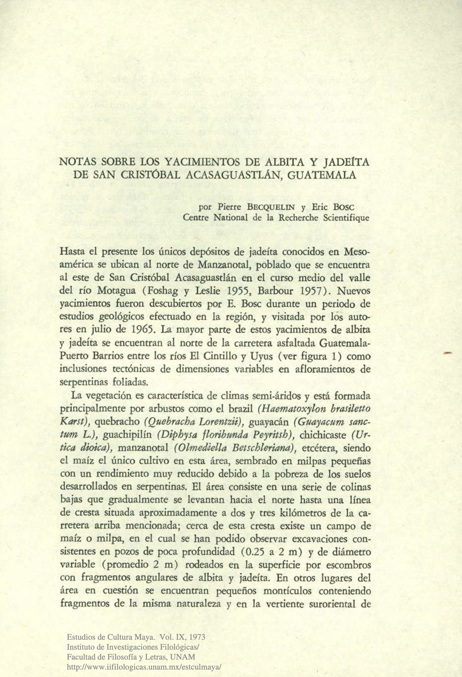
Notas sobre los yacimientos de albita y jadeita de San Cristobal Acasaguastlan, Guatemala
Notes on the albite and jadeite deposits of San Cristóbal Acasaguastlán, Guatemala
Journal Article
Becquelin, P. and Bosc, E. (1973) Notas sobre los yacimientos de albita y jadeita de San Cristobal Acasaguastlan, Guatemala. [Notes on the albite and jadeite deposits of San Cristóbal Acasaguastlán, Guatemala] Estudios de Cultura Maya, Vol. 9, pp. 67–73.
Record No. 28417  |  Sinkankas/Born No.: NIS/NIB  |  Language: in Spanish  |  RWHL
Up to the present, the only jadeite deposits known in Mesoamerica are located north of Manzanotal, a settlement situated east of San Cristóbal Acasaguastlán in the middle course of the Motagua River Valley (Foshag and Leslie 1955; Barbour 1957). New deposits were discovered by E. Böse during a period of geological studies carried out in the region and were visited by the authors in July 1965. Most of these albite and jadeite deposits are located north of the paved Guatemala–Puerto Barrios highway, between the El Cintillo and Uyús rivers (see Figure 1), occurring as tectonic inclusions of varying dimensions within outcrops of foliated serpentinite. Becquelin and Bosc describe newly identified albite and jadeite occurrences near San Cristóbal Acasaguastlán in Guatemala’s middle Motagua Valley, where the deposits occur as tectonic inclusions of varying dimensions within foliated serpentinite. The authors report shallow pits surrounded by angular fragments of albite and jadeite, small mounds, terraces, and nearby obsidian blade fragments, which together suggest rudimentary extraction, possibly in prehistoric times, although they stress that further investigation is required to establish the nature and age of the workings. Other jadeite occurrences in the region lack evidence of ancient exploitation. The archaeological evidence from nearby Guaytán, Manzanotal, and other middle Motagua sites includes jade fragments, partly worked beads and drills, and a jadeite block bearing percussion marks. The authors suggest that inhabitants of the valley collected albite and jadeite from terraces and gravels of the Motagua and perhaps directly from serpentinite sources, manufactured beads and other simple ornaments, and exported jade as blocks and, in some cases, finished objects. They conclude that exploitation and trade in jade probably flourished during the Classic period and that the San Cristóbal region may have supplied a substantial portion of the jade used elsewhere in Mesoamerica.

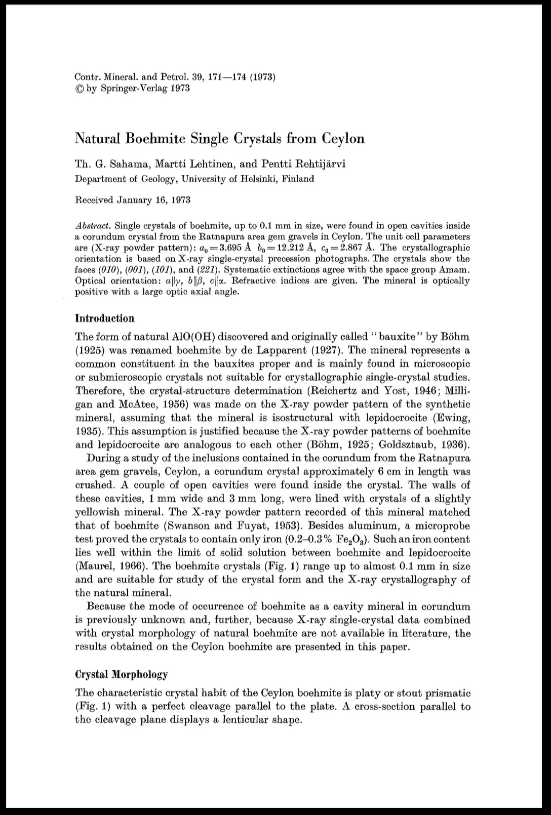
Natural boehmite single crystals from Ceylon
Journal Article
Sahama, T.G., Lehtinen, M. and Rehtijärvi, P. (1973) Natural boehmite single crystals from Ceylon. Contributions to Mineralogy and Petrology, Vol. 39, pp. 171–174.
Record No. 4959  |  Sinkankas/Born No.: NIS/NIB  |  RWHL
Natural boehmite single crystals approaching 0.1 mm in size are described from open cavities within a corundum crystal recovered from the gem gravels of the Ratnapura area, Ceylon. The occurrence provides material suitable for relating the morphology, crystallographic orientation, and optical properties of natural boehmite, which generally occurs in crystals too small for single-crystal investigation. Crushing a corundum crystal approximately 6 cm long exposed cavities about 1 mm wide and 3 mm long, lined with slightly yellowish crystals. Identification rests on an X-ray powder pattern matching boehmite, supported by microprobe examination showing aluminium and only minor iron, equivalent to 0.2–0.3 per cent Fe₂O₃. The crystals are platy or stout prismatic, possess perfect cleavage parallel to the plate, and exhibit a lenticular outline in sections parallel to that cleavage. The study resolves inconsistencies in earlier descriptions caused by differing choices of crystallographic axes, adopting the conventional setting in which b is longer than a and a longer than c. Single-crystal precession photographs establish the orientation, while microscopic measurements of interfacial angles permit identification of the forms (010), (001), (101), and (221). Unlike previously described synthetic plates, the Ceylon crystals develop (001) rather than (100), and no cleavage parallel to (100) is observed. Powder diffraction yields unit-cell dimensions of a = 3.695 ± 0.003 Å, b = 12.212 ± 0.006 Å, and c = 2.867 ± 0.003 Å. Systematic extinctions agree with the orthorhombic space group Amam, although the authors retain the possibility that asymmetric hydrogen bonding makes the true structure noncentrosymmetric. Optical orientation is a parallel to γ, b parallel to β, and c parallel to α. Immersion measurements give refractive indices α = 1.648, β = 1.657, and γ = 1.668, each with an uncertainty of ±0.002. Because these uncertainties limit calculation of the optic axial angle, birefringence is measured independently with a Berek compensator, yielding γ–α = 0.020 and γ–β = 0.012. These values indicate a large optic axial angle of approximately 79° and positive optical character, supported by qualitative conoscopic observations. The paper documents a previously unreported mode of occurrence and provides coordinated morphological, X-ray, and optical data for natural boehmite without proposing a formation mechanism for the cavity crystals.

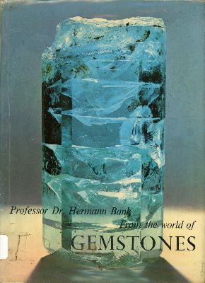
From the World of Gemstones

Book
Bank, H. (1973) From the World of Gemstones. Innsbruck, Pinguin-Verlag, 178 pp.
Record No. 422  |  Sinkankas/Born No.: NIS/NIB  |  RWHL
Bank presents an illustrated survey of gemstones, introducing their chemical and physical properties and explaining how these characteristics relate to identification. Individual gem materials are arranged according to mineralogical classification, linking their descriptions to the broader study of minerals. The text discusses inclusions and other diagnostic features, while locality maps provide geographical context for the occurrences described. Colour photographs form a major part of the presentation, allowing readers to appreciate the appearance and variety of gem materials alongside the scientific account. The work combines introductory explanations with information intended for readers already familiar with gemmology, serving as a general reference and visual survey. Its treatment of testing is selective, with less emphasis on absorption spectroscopy, and the individual descriptions do not consistently supply refractive-index and specific-gravity values. E.H. Rutland’s English translation makes Bank’s German account accessible to an English-speaking readership.

Developments and highlights at GIA’s lab in New York: Glass
Journal Article
Crowningshield, G.R. (1973) Developments and highlights at GIA’s lab in New York: Glass. Gems & Gemology, Vol. 14, No. 5, Spring, pp. 134–137.
Record No. 138  |  ISBN/ISSN: 0016626X  |  Sinkankas/Born No.: NIS/NIB  |  RWHL
Crowningshield reports several glass gemstone imitations examined at GIA’s New York laboratory, emphasizing how convincing colour, internal crystallization, and manufactured structures can complicate identification. A group of green jade substitutes submitted within a few weeks ranged from ordinary translucent glass to partly devitrified material closely resembling fine fei cui. One relatively straightforward imitation was mounted in platinum with diamonds, illustrating that an elaborate setting does not establish the identity of its principal stone. Another fine green example showed an unusual radiating fibrous pattern. Crowningshield relates such crystalline or partly crystalline structures in glass to devitrification and compares them with the fern-leaf appearance of the Japanese imitation sold as meta-jade or Iimori stone. In that ring, the apparent surrounding diamonds and their prongs were simulated by cutting the metal setting itself. A further 31 ct cabochon resembled valuable imperial fei cui but contained irregular crystal-like structures or tufts produced by devitrification. Another imitation was identifiable as glass from prominent gas bubbles, although its refractive index of 1.66 and mottled appearance so closely approached those of fei cui that two trade members wagered on the laboratory’s conclusion. These cases demonstrate that apparently mineral-like inclusions and plausible optical measurements must be considered alongside decisive microscopic evidence. Other glass substitutions included a bracelet purchased as natural emerald crystals, whose components were manufactured hexagonal prisms, mostly green and sometimes incorporating a brown imitation matrix. The report also describes attempts to reproduce cat’s-eye phenomena in glass. An early white product contained visible hollow tubes that scattered light and generated chatoyancy, while later imitations were available in several colours, including honey-like shades and pale blue. Some displayed sharp eyes despite structures too fine to resolve readily at ordinary gemmological magnification. Side views revealed honeycomb patterns, initially attributed tentatively to devitrification. A brownish yellow example resembling chrysoberyl showed a pronounced fibre-optic effect and larger hexagonal areas composed of numerous smaller hexagonal fibres. Its precise manufacturing method remained uncertain, although fused optical fibres were suggested; its refractive index exceeded the instrument’s scale and its specific gravity was 4.19. Photographs document the internal textures and viewing effects. Collectively, the observations show the variety of glass substitutes and the importance of examining structural details from several directions rather than relying on colour, apparent inclusions, jewellery quality, or a single test.

On the occurrence of Chlorapatite at the Emerald Mine, Rajagarh village, Ajmer
Journal Article
Babu, S.K. (1972) On the occurrence of Chlorapatite at the Emerald Mine, Rajagarh village, Ajmer. Mineralogical Magazine, Vol. 38, No. 300, December, pp. 972–974.
Record No. 27262  |  ISBN/ISSN: 0026461X  |  DOI: https://doi.org/10.1180/minmag.1972.038.300.10  |  Sinkankas/Born No.: NIS/NIB  |  RWHL
S.K. Babu describes the occurrence, optical properties, chemistry, X-ray diffraction characteristics, and inferred origin of yellowish-green apatite collected at the emerald mine near Rajagarh village, Ajmer, India. The crystals, initially mistaken for emerald, occur at the contact between mica schist and pegmatite within a terrain of schists and gneisses associated with altered peridotites and intrusive granite. Described as chlorapatite or asparagus stone, the material is identified more specifically in the diffraction table as chlorian hydroxyapatite. The crystals are semitransparent, with vitreous lustre, uneven to subconchoidal fracture, and a specific gravity of 3.17. Preparation for analysis involved crushing a crystal and separating gangue with diluted Clerici’s solution, yielding approximately 99% pure apatite. Minute sericite inclusions remained and were estimated by point counting to constitute 0.93% of the separated material. In thin section the apatite is colourless and contains acicular inclusions and possible fluid inclusions. It is optically negative but displays a pseudo-biaxial figure, attributed tentatively to strain. Immersion measurements give refractive indices of 1.642 and 1.649, slightly above those of common apatite and comparable with published chlorapatite values. Powder diffraction spacings and intensities likewise compare favourably with reference data; the pattern is indexed using unit-cell parameters a = 9.598 Å and c = 6.739 Å. Chemical analysis combines gravimetric, volumetric, flame-photometric, and atomic-absorption methods, with water determined by ignition with calcium oxide. The reported composition includes 41.50% phosphorus pentoxide, 55.69% calcium oxide, 0.45% fluorine, 1.31% chlorine, and 0.73% water released on ignition, together with minor constituents and the sericite impurity. Trace-element investigation is directed toward distinguishing an igneous or pegmatitic origin from a metamorphic one. Comparison with published apatite analyses shows a distribution resembling that of granitic pegmatites, particularly relatively high manganese, yttrium predominating over cerium, and low strontium. A plot of cerium against silicon also places the specimen within the published field for pegmatitic apatites. Babu therefore concludes that the mineral belongs genetically to the granitic pegmatite rather than to the surrounding metamorphic rocks, combining its contact-zone occurrence with optical, structural, and geochemical evidence.

Inclusions in gemstones

Journal Article
Gübelin, E.J. (1972) Inclusions in gemstones. Australian Gemmologist, Vol. 11, No. 8, November, pp. 3–14.
Record No. 893  |  Sinkankas/Born No.: NIS/NIB  |  RWHL

Sapphire substitutes

Journal Article
Anderson, B.W. (1972) Sapphire substitutes. Journal of Gemmology, Vol. 13, No. 1, January, p. 4.
Record No. 242  |  ISBN/ISSN: 221252  |  Sinkankas/Born No.: NIS/NIB  |  RWHL
Anderson reports two unusual sapphire substitutes, emphasizing the identification difficulties presented by a composite combining natural and synthetic sapphire. He places these observations against a background in which familiar substitutes included Verneuil synthetic sapphires, garnet-topped doublets, and glass imitations, while the appearance of blue zoisite had introduced another material requiring consideration. The first unexpected example was a step-cut agate, dyed blue with greenish stripes and mounted in a cluster ring. Its banding gave a surprisingly convincing impression of a natural sapphire of poor quality, and an experienced dealer had bought and sold it in good faith under that description. The principal concern is a more sophisticated doublet apparently being marketed in appreciable quantities. A green or greenish crown of natural Australian sapphire is joined to a blue synthetic sapphire base, with the junction positioned just below the girdle. This joining line is difficult to detect with a lens even when the stone is loose and would be concealed by mounting. Parcels had been offered at approximately £50–£60 per carat, prices consistent with their apparent value as natural sapphires, and several examples had initially been accepted as genuine. The construction is particularly deceptive because examination through the table reveals natural inclusions in the crown, while spectroscopy clearly shows the absorption complex near 450 nm that had previously been considered sufficient evidence of natural origin. These observations therefore establish the natural character of part of the composite without demonstrating that the entire stone is natural. In the specimens examined, immersion and inspection in profile exposed the construction readily. The lower portions displayed gas bubbles and curved growth bands characteristic of Verneuil synthetic sapphire. They also showed whitish fluorescence under short-wave ultraviolet radiation, a feature Anderson identifies as potentially useful when examining mounted examples. He qualifies the ease of these identifications by noting that the specimens were loose and that the examiners had been forewarned. The report warns that apparently persuasive inclusions and absorption features can be misleading when natural material forms only the crown of an assembled stone, making thorough examination of the pavilion and possible junction essential.

Notes from the laboratory: Kashan synthetic rubies; ruby doublets
Journal Article
Anderson, B.W. (1972) Notes from the laboratory: Kashan synthetic rubies; ruby doublets. Journal of Gemmology, Vol. 13, No. 3, July, pp. 96–97.
Record No. 241  |  ISBN/ISSN: 221252  |  Sinkankas/Born No.: NIS/NIB  |  RWHL
Describes the identifying features of Kashan synthetic rubies and a type of ruby doublet combining natural sapphire with synthetic ruby. Rough and faceted Kashan specimens lent by M.J. O’Donoghue were examined against the background of their reputation and relatively high price. These rubies were grown from a flux melt at normal atmospheric pressure and were reportedly no longer in production at the time of writing. The rough crystals differed from natural ruby in habit, forming flattened rhombohedra with partially resorbed surfaces. Their subdued red colour, weak absorption spectrum, and weak fluorescence spectrum indicated a surprisingly low chromium content. Microscopic examination revealed the characteristic flat flux inclusions previously described by Gemological Institute of America investigators as resembling paint splashes, although these occurred on a relatively small scale in the specimens examined. Individual inclusions were elongated, with parallel sides and rounded ends, and occurred in parallel groups. Twisted, lace-like feathers characteristic of many flux-grown stones were also present. Anderson particularly noted the absence of included foreign minerals. The second note extends earlier observations on doublets comprising a natural Australian sapphire crown and a synthetic sapphire base. In the newer construction, a natural sapphire crown was combined with a synthetic ruby base to produce an effective ruby imitation. The blue or greenish natural upper component supplied natural silk, while the red synthetic base dominated the overall colour sufficiently to mask the crown’s blue-green contribution. This combination could therefore present both the colour expected of ruby and inclusions associated with natural corundum. Differential fluorescence provided an important means of detection: under crossed filters or ultraviolet illumination, the natural sapphire crown remained non-fluorescent while the synthetic ruby base glowed bright red. Microscopic examination also revealed an appearance inconsistent with an entirely natural stone. Anderson warned that these composites could deceive many observers unless their construction and identifying characteristics became widely recognised. Together, the notes emphasise the value of examining crystal habit, inclusion morphology, spectral behaviour, and the separate optical responses of a composite stone’s components when distinguishing synthetic and assembled ruby substitutes.

Mafic and ultramafic inclusions associated with undersaturated basalt on Kod Ali Island, Southern Red Sea
Journal Article
Hutchison, R. and Gass, I.G. (1971) Mafic and ultramafic inclusions associated with undersaturated basalt on Kod Ali Island, Southern Red Sea. Contributions to Mineralogy and Petrology, Vol. 31, No. 2,, 6/1/71 pp. 94–101.
Record No. 17256  |  ISBN/ISSN: 14320967  |  DOI: https://doi.org/10.1007/BF00373452  |  Sinkankas/Born No.: NIS/NIB  |  RWHL
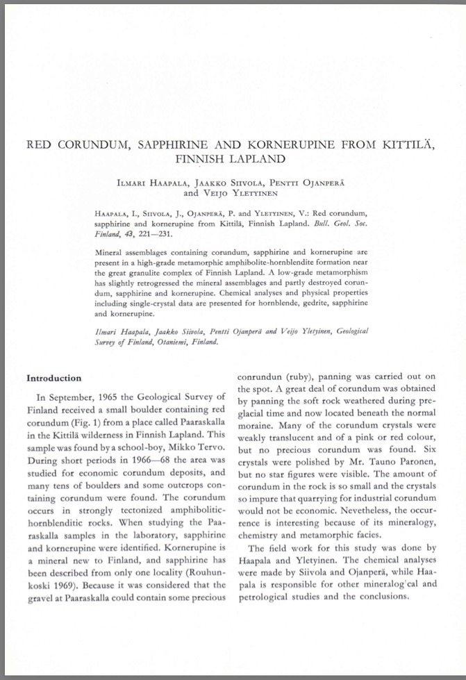
The island of Kod Ali is a small diatreme cone lying close to the Ethiopian coast at latitude 13° 57′ N, longitude 41° 49′ E. This volcanic structure, produced by geologically Recent volcanic activity, consists, almost entirely, of pyroclastic debris. Evidence of a liquid phase is confined to the thin veneers of markedly alkalic olivine basalt which coat many of the abundant gabbroic, pyroxenitic and lherzolitic fragments found on the island. As silicic inclusions are completely absent and as the alkalic liquid phase and the “tholeiitic” inclusions are seemingly genetically unrelated, it is proposed that the latter might have been derived from a tholeiitic plutonic layered sequence that underlies the tholeiitic basalts of an “oceanic” Red Sea floor.

Red corundum, sapphirine and kornerupine from Kittilä, Finish Lappland
Journal Article
Haapala, I., Siivola, J., Ojanpera, P. and Yeletinen, V. (1971) Red corundum, sapphirine and kornerupine from Kittilä, Finish Lappland. Bulletin, Geological Society of Finland, Vol. 43, pp. 221–231.
Record No. 7642  |  DOI: https://doi.org/10.17741/bgsf/43.2.007  |  Sinkankas/Born No.: NIS/NIB  |  RWHL
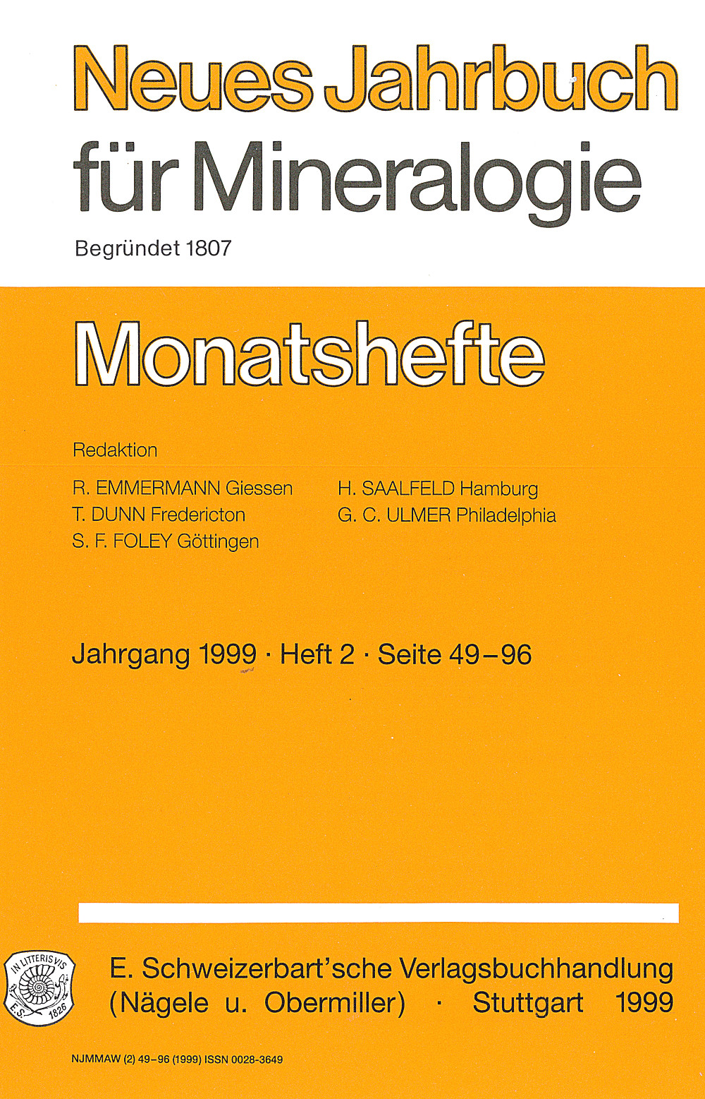
Corundum-, sapphirine- and kornerupine-bearing rocks from Paaraskalla near Kittilä in Finnish Lapland are described through field observations, petrography, chemical analyses and crystallographic measurements. Investigation followed the discovery of a red-corundum-bearing boulder in 1965 and located numerous additional boulders and several outcrops within a strongly deformed amphibolite–hornblendite formation near Lapland’s major granulite complex. Corundum-bearing material follows a narrow zone broadly parallel to the formation’s foliation. The principal rock-forming minerals are hornblende and plagioclase, with distinctive assemblages containing corundum, gedrite, sapphirine or kornerupine. Corundum is generally pink, white or red, although small deep blue grains were also observed. Crystals commonly measure about one centimetre or less, with occasional larger examples. Panning of weathered rock beneath the moraine yielded abundant corundum, but no gem-quality material was recovered, six polished specimens showed no asterism, and the low concentration and impurity of the crystals made industrial extraction uneconomic. The occurrence is therefore significant chiefly for its mineralogy and metamorphic history. Detailed chemical compositions, optical properties, densities and single-crystal data are presented for hornblende, gedrite, sapphirine and kornerupine. The hornblende is magnesium- and aluminium-rich, while the sapphirine has unusually high alumina and relatively low silica compared with previously analysed examples. Pale bluish green kornerupine, recognised only in the original boulder, represents a new mineral occurrence for Finland and contains relatively little boron. Mineral textures document substantial replacement: corundum and sapphirine are partly converted to chlorite, kornerupine is strongly altered to chlorite and fine mica, and exposed anorthite is replaced by sericite and clinozoisite while shielded inclusions remain fresh. Dravite associated with corundum suggests boron metasomatism. The assemblages are interpreted as products of high-pressure, high-temperature metamorphism near the amphibolite–granulite facies boundary, followed by greenschist-facies retrogression that partly destroyed the diagnostic minerals. Experimental phase-equilibrium results provide comparisons, but the authors caution against transferring them directly to these chemically complex natural rocks. Whole-rock chemistry, accessory chromite and possible replacement of former pyroxene by hornblende favour a mafic–ultramafic igneous precursor, potentially with aluminium- and magnesium-rich layers. Poor exposure and limited evidence nevertheless prevent exclusion of a metasedimentary origin.

Korundeinschlüsse im Korund

Corundum inclusions in corundum
Journal Article
Eppler, W.F., Jagodzinski, H. and Oppermann, J. (1971) Korundeinschlüsse im Korund. [Corundum inclusions in corundum] Neues Jahrbuch für Mineralogie, Monatshefte, No. 10, pp. 429–432.
Record No. 7623  |  Sinkankas/Born No.: NIS/NIB  |  Language: in German  |  RWHL
Eppler, Jagodzinski, and Oppermann describe microscopic inclusions of corundum occurring within corundum crystals and examine their crystallographic relationships with the host. The study investigates the orientation and structural relationship of the included corundum crystals, using crystallographic observations to determine the nature of their intergrowth with the surrounding host. The authors discuss the significance of these inclusions for understanding corundum growth and crystallization, showing that inclusions of the same mineral species as the host can preserve evidence of distinct stages or conditions of crystal development.

Exploring the Literature of Gemstone Inclusions

The literature of gemstone inclusions covers identification, origin determination, treatments, synthesis, mineral inclusions, fluid inclusions and other internal features of natural and synthetic gems.

For a curated selection of important sources, read Inclusions in Gemstones — Hyperion Literature Sources.

About Four Treasures

Four Treasures is Lotus Gemology's freely accessible gemological reference database. Use the full database to search by author, title or keyword and explore literature on gemology, mineralogy, jewelry and related fields. Where available, hyperlinks on titles may lead directly to the full publication.

Search the Complete Four Treasures Database