Gemstone Inclusions Bibliography

Gemstone Inclusions Bibliography

Explore books, articles and historical sources on inclusions in gemstones in Four Treasures, the Lotus Gemology reference database. This bibliography brings together matching records from the collection, with publication details, abstracts and cover images where available.

Gemstone Inclusion References

The list below matches the terms inclusion or inclusions in the database's searchable fields. Some references discuss inclusions as part of a broader subject.

941–960 of 1,344 matching references

Borsen Bulletin: Rubies from Vietnam

Journal Article
Bank, H. and Henn, U. (1990) Borsen Bulletin: Rubies from Vietnam. Goldschmiede und Uhrmacher Zeitung, No. 12,, December p. 106.
Record No. 4703  |  Sinkankas/Born No.: NIS/NIB  |  RWHL

Die brasilianischen Smaragde und ihre Vorkommen: Santa Terezinha de Goiás/GO.
The brazilian emeralds and their deposits: Santa Terezinha de Goiás, GO
Journal Article
Schwarz, D. (1990) Die brasilianischen Smaragde und ihre Vorkommen: Santa Terezinha de Goiás/GO. [The brazilian emeralds and their deposits: Santa Terezinha de Goiás, GO] Zeitschrift der Deutschen Gemmologischen Gesellschaft, Vol. 39, No. 1, pp. 13–44.
Record No. 4200  |  Sinkankas/Born No.: NIS/NIB  |  Language: In German  |  RWHL
Describes the emerald deposits of Santa Terezinha de Goiás, Brazil, presenting evidence for their stratiform occurrence and documenting the stones’ gemological properties, chemistry and inclusions. Refractive indices reach 1.593–1.602, with birefringence of 0.008–0.010, reported as the highest values then observed in emerald. Chromium and iron show the highest mean concentrations among Brazilian emeralds. Fluid inclusions are generally very small. Mineral inclusions include spinel, carbonates, mica, talc, amphibole and pyrite, with rutile, hematite, chalcopyrite, pentlandite and ilmenite also recorded.

Lab Notes: Cobalt-colored color-change spinel
Journal Article
Maddison, P. (1990) Lab Notes: Cobalt-colored color-change spinel. Gems & Gemology, No. 3, Fall, pp. 225–226.
Record No. 4142  |  ISBN/ISSN: 0016626X  |  Sinkankas/Born No.: NIS/NIB  |  RWHL
A laboratory examination documents an unusual colour change in a 2.24-carat natural cobalt-coloured spinel mounted in a man’s ring. The stone appeared dark, richly saturated, slightly violetish blue under fluorescent illumination and dark purple under incandescent light. Its contemporary setting, combining white and yellow metals, enhanced the visual effect. Testing at the West Coast laboratory established properties consistent with natural cobalt-blue spinels examined previously, including examples reportedly originating in Sri Lanka, although the note does not establish a geographic source for this particular specimen. A Duplex II refractometer yielded a single refractive-index reading of 1.720. Polariscope examination showed a singly refractive response accompanied by very slight anomalous double refraction. The stone displayed weak red transmission and appeared moderately red through a Chelsea colour filter. Examination with a handheld spectroscope revealed the diagnostic absorption band of natural cobalt spinel at approximately 460 nm, together with additional bands near 480, 552, 559, 575, and 622 nm. Broad general absorption extended from approximately 660 nm towards longer wavelengths. These optical observations supported comparison with the laboratory’s earlier experience of natural cobalt-blue spinel despite the specimen’s unusual change between blue and purple. Microscopic examination beneath one end of the crown revealed a cluster of moderately sized inclusions comprising a well-formed octahedral crystal and several whitish crystals of irregular shape. Rain-like strings of pinpoint inclusions radiated from this group and extended through the stone, accompanied by numerous small, fine needles. The included crystals and needles are described by their appearance and morphology without assignment to specific mineral species. The report combines observations under different light sources with refractometry, polariscope testing, colour-filter examination, absorption spectroscopy, and microscopy to characterize the mounted gemstone. Its significance lies in documenting a distinctive blue-to-purple colour change in a stone whose other measured properties and internal features were consistent with natural cobalt-blue spinel.

Gem Trade Lab Notes: Spinel from Tanzania
Journal Article
Kane, R.E. (1990) Gem Trade Lab Notes: Spinel from Tanzania. Gems & Gemology, Vol. 26, No. 2, Summer, pp. 156–157.
Record No. 4135  |  ISBN/ISSN: 0016626X  |  Sinkankas/Born No.: NIS/NIB  |  RWHL
A 2.28 ct purplish pink faceted pear-shaped stone submitted to the West Coast laboratory was identified as natural spinel and reported by its owner to originate from Tanzania. Its refractive index was 1.714, and it was singly refractive with very weak anomalous double refraction. Hydrostatic specific gravity was approximately 3.60. The stone displayed strong red fluorescence under long-wave ultraviolet radiation, weak red fluorescence under short-wave radiation, and a characteristic absorption spectrum. Its most unusual optical feature was a moderate to strong whitish blue transmission effect observed when intense light was directed through a small opening onto the stone. The phenomenon was also visible without magnification when the spinel was held a few inches from an incandescent or fluorescent source, while a faint bluish haze could be seen at arm’s length in sunlight. The effect was interpreted as probably resulting from light scattering by minute inclusions or dislocations, comparable to similar reactions in other gem materials. Microscopic examination showed that the stone was only slightly included. Parallel rows of tiny octahedra, commonly encountered in spinels from classic Myanmar and Sri Lankan localities, were conspicuously absent. Instead, the dominant internal features were numerous sheets of polysynthetic twinning intersecting in two directions throughout the gem. Coarse needles or growth tubes were associated with these planes in several areas. Immersion in methylene iodide revealed faint color zoning apparently aligned with some of the twinning features. Densely packed clouds of minute particles occurred through much of the stone, appeared to represent exsolutions, and were oriented in three directions. A few small, thin, transparent, nearly colorless tabular euhedral crystals were also present. The combination of physical and optical properties, color, and visual appearance matched spinels attributed to a newly reported Tanzanian deposit that the author had examined during a visit to Idar-Oberstein. Tanzanian spinel was also available at the 1990 Tucson Show. The note documents distinctive optical and microscopic characteristics of this specimen while retaining its Tanzanian origin as a reported attribution.

Crystals in ruby

Journal Article
Themelis, T. (1990) Crystals in ruby. Lapidary Journal, Vol. 43, No. 11, February, p. 19.
Record No. 1713  |  Sinkankas/Born No.: NIS/NIB  |  RWHL

Multiple chatoyancy in sapphire

Journal Article
Pearson, G. (1990) Multiple chatoyancy in sapphire. Australian Gemmologist, Vol. 17, No. 8, pp. 296–298.
Record No. 1413  |  Sinkankas/Born No.: NIS/NIB  |  RWHL
Unusual multiple chatoyancy in Australian sapphire is investigated through visual and microscopic examination of cabochons reportedly cut from Rubyvale, Queensland, and Inverell, New South Wales, rough. The study began with a Rubyvale sapphire measuring 10 × 8 × 4.5 mm that displayed a weak, diffuse white star together with three bright, mobile satellite reflections on its dome. Examination of two parcels revealed several comparable Rubyvale specimens, including four stones with three symmetrically arranged satellite reflections and three small twelve-rayed star sapphires; one specimen combined both features. The Inverell parcel yielded no comparable multiple chatoyancy, although one small stone displayed two stars attributed to twinning. Ordinary asterism is explained by reflection from crystallographically oriented acicular inclusions, whereas the additional reflections are associated with aligned, approximately equant platelets whose individual reflections appear as spots rather than lines. These inclusions occur predominantly in bands approximately parallel to prism faces, and rough angular measurements suggest reflecting surfaces inclined about 45° to the basal plane. Their mineral identity remains uncertain: non-acicular rutile, anatase and titanite are considered possible constituents. Most multiply chatoyant specimens were deep blue, strongly zoned with silk and green when viewed perpendicular to the crystal axis, features taken to suggest appreciable iron content. Their satellite reflections occupied alternate arms of a bronze-coloured star. A pale-blue exception displayed a diffuse white star with the three reflections positioned between its arms. Detailed examination of a twelve-rayed specimen identified two inclusion systems producing white and bronze six-rayed stars rotated approximately 30° relative to each other. The white system consisted of fine, long, relatively colourless needles resembling rutile, while the bronze system comprised shorter, tapering, blade-like inclusions concentrated in bands associated with hexagonal or chevron zoning. Some bronze blades showed interference colours consistent with very thin platelets. An iron-rich, titanium-depleted ilmenite-related phase, discussed in connection with previous work on ferrilmenite, is proposed for the bronze silk. The article documents the geometrical relationships between the stars, satellite reflections and host-crystal symmetry, while presenting the identities and formation mechanisms of the responsible inclusions as hypotheses requiring further confirmation.

Crystals

Book
Mercer, I.F. (1990) Crystals. Cambridge, MA, Harvard University Press, 60 pp.
Record No. 1257  |  Sinkankas/Born No.: NIS/NIB  |  RWHL
An illustrated introduction to crystals explains how their internal atomic order governs external form, growth, physical properties, and practical uses. Photographs, diagrams, and examples from minerals, gemstones, household substances, living organisms, and manufactured materials connect the microscopic structure of crystals with familiar objects. Repeating atomic arrangements are introduced through comparisons with patterned surfaces and three-dimensional building blocks, leading to unit cells, lattice types, symmetry elements, crystal systems, and crystal classes. The distinction between crystal form and habit is explained, emphasising that unequal growth may distort outward shape without changing the characteristic angles between corresponding faces. Cleavage, twinning, surface markings, colour zoning, and inclusions are presented as visible expressions of structure or records of growth history. The geological discussion relates crystals to rock formation, the Earth’s composition, magmas, mineral-bearing solutions, metamorphism, and extraterrestrial materials. Crystal growth is described in liquids, solids, and vapours, including nucleation, saturation, cooling, evaporation, recrystallisation, and changes in stability under different temperatures and pressures. Simple activities demonstrate growing crystals from solution, melt, and vapour. A recurring theme is that real crystals are imperfect: dislocations, impurities, intergrowths, and other defects may influence growth rates, deformation, colour, and optical effects, often producing useful properties rather than merely flaws. Interactions with light and X-rays introduce polarisation, double refraction, pleochroism, diffraction, and methods of identifying minerals and determining structure. Practical sections describe observation with a hand lens, measurement of face angles, simple optical instruments, and approaches to collecting specimens, pebbles, micromounts, and gems. Natural gem materials are considered alongside synthetic crystals, with examples of flame-fusion growth, crystal pulling, hydrothermal quartz production, flux-grown emerald, and high-pressure diamond synthesis. Applications range from abrasives and construction materials to quartz watches, lasers, semiconductors, light-emitting diodes, and molecular sieves. Brief treatments of crystal lore and the history of crystallography place modern structural understanding in a wider context. Reference tables, notable crystal specimens, size records, and suggested reading supplement a guide that encourages direct observation and experimentation while linking crystal beauty to the behaviour of ordered matter.

The most common misconceptions about flux grown rubies
Journal Article
Carter, V.L. (1990) The most common misconceptions about flux grown rubies. AGA Cornerstone, July, pp. 39–41.
Record No. 554  |  Sinkankas/Born No.: NIS/NIB  |  RWHL
Discusses seven misconceptions about flux-grown synthetic rubies from the perspective of a partner in the J.O. Crystal Company, producer of Ramaura ruby. The author first rejects the assumption that laboratory growth permits unlimited control over crystal size, clarity, variety and cost, stressing that synthesis remains constrained by physical laws and practical growth conditions. Inclusions are not deliberately inserted to imitate natural ruby: trapped flux, internal fractures, fingerprints and feathers arise during growth, and the producer seeks to minimise them because cleaner material commands higher prices. The article distinguishes the implications of the terms laboratory-grown, flux-grown, created and cultured, explaining the company’s preference for “cultured” as a description of crystals grown from a nutrient solution with human involvement. It contrasts flux growth with inexpensive, high-volume flame-fusion production and argues that the two products differ substantially in appearance and manufacturing cost. Carter attributes Ramaura ruby’s natural-looking appearance to slow precipitation from a supersaturated solution, spontaneous nucleation without a seed, and a relatively regular crystal lattice with few dislocations. Addressing fears that synthetic stones will overwhelm the market, she states that annual flux-grown ruby and emerald production amounts to no more than approximately 1% of the number of newly mined and faceted natural counterparts, and that difficult, costly production prevents supply from meeting demand. Particular attention is given to the identification dopant intentionally incorporated into every marketed Ramaura ruby. According to the author, this shifts long-wave ultraviolet fluorescence towards yellow-orange relative to natural ruby; its effect varies with the dopant concentration prevailing when individual crystals form. Examination should therefore take place in a darkened room, using a long-wave ultraviolet lamp and direct comparison with a natural ruby. The company describes doping as a voluntary measure intended to preserve reliable distinction from natural material. Finally, the article explains why synthetic origin does not imply negligible value: high-purity platinum crucibles, months of furnace operation, substantial electricity consumption and low cutting yields all contribute to cost. Reported prices range from US$50–350 per carat according to size and grade, or approximately 5–10% of the price of comparable natural ruby. Company and partner biographies provide context for these explanations and the commercial position from which they are presented.

Thermal alteration of inclusions in ‘rutilated’ topaz,
Journal Article
Kammerling, R.C. and Koivula, J.I. (1989) Thermal alteration of inclusions in ‘rutilated’ topaz,. Gems & Gemology, Vol. 25, No. 3, Fall, pp. 165–167.
Record No. 26771  |  Sinkankas/Born No.: NIS/NIB  |  RWHL
Colorless faceted topaz containing acicular inclusions has been marketed as “rutilated” topaz, though earlier studies revealed that these inclusions are actually limonite-stained etched dislocation channels rather than rutile. This article discusses the application of heat treatment to convert the limonite staining into hematite, thereby enhancing the visibility and altering the color of the inclusions to make them appear more prominent.

Spinelle aus dem Umba-Tal, Tanzania

Spinels from the Umba Valley, Tanzania
Journal Article
Bank, H., Henn, U. and Petch, E. (1989) Spinelle aus dem Umba-Tal, Tanzania. [Spinels from the Umba Valley, Tanzania] Zeitschrift der Deutschen Gemmologischen Gesellschaft, Vol. 38, pp. 166–168.
Record No. 5988  |  Sinkankas/Born No.: NIS/NIB  |  Language: In German  |  RWHL
A brief gemological report on spinels from Tanzania’s Umba Valley, occurring in pink, red, orangish red and bluish violet colors. The specimens examined had refractive indices of 1.713–1.718 and specific gravities of 3.54–3.69. Spectroscopic examination was used to investigate the causes of the different colors. The note documents the range of gem spinel then reaching the market from the Umba Valley and provides basic properties useful for characterizing this East African material.

Gem News: An interesting zoned Australian sapphire
Journal Article
Koivula, J.I. and Kammerling, R.C. (1989) Gem News: An interesting zoned Australian sapphire. Gems & Gemology, Vol. 25, No. 2, Summer, p. 114.
Record No. 5403  |  ISBN/ISSN: 0016626X  |  Sinkankas/Born No.: NIS/NIB  |  RWHL

Star sapphire from Kenya

Journal Article
Gübelin, E.J., Barot, N.R., Flamini, A. and Graziani, G. (1989) Star sapphire from Kenya. Journal of Gemmology, Vol. 21, No. 8, October, pp. 467–473.
Record No. 5066  |  ISBN/ISSN: 221252  |  Sinkankas/Born No.: NIS/NIB  |  RWHL
Examines cabochon-cut sapphires from northwestern Kenya to characterise their inclusions, explain their asterism and distinguish them from synthetic star sapphires. Material entering the Kenyan market from mid-1987 was collected from superficial secondary deposits west of Lake Turkana, although its primary geological source remained unidentified. Ten cabochons, ranging from blue and greenish blue to greenish yellow and dark brown, were investigated using conventional gemmological testing, optical microscopy, electron-microprobe analysis and X-ray diffraction. Eight displayed six- or twelve-rayed asterism of varying quality, while two showed concentrated silky areas rather than a complete star. Some specimens produced exceptionally sharp, evenly developed six-rayed stars resembling those of Linde synthetic sapphires. Refractive-index measurements gave an approximate value of 1.765, and the stones were inert under long- and short-wave ultraviolet radiation. Visible absorption commonly showed iron-related features near 450, 460 and 470 nm. Microscopic examination documented cloudy areas, acicular structures, rutile silk, tubular cavities, transparent crystals, fluid feathers and healed fractures. The structures associated with asterism were separated into microstriae compositionally similar to the sapphire host and distinct needle-like inclusions interpreted as rutile or hematite. Their crystallographically controlled orientations and variable distribution were considered responsible for differences in star development. Rutile was supported by weak reflections in bulk X-ray diffraction patterns. Darker colours were associated with iron-rich inclusions, including hematite needles and iron oxide or hydroxide material occupying channels and healed fractures. Transparent, low-relief hexagonal crystals were assigned to corundum, while reddish-brown prismatic titanium-rich inclusions were interpreted as rutile. Straight-edged colour bands and angular growth zoning record successive growth episodes and changing chemical conditions. Together with the absorption spectrum, these features help distinguish the natural material from synthetic star sapphire despite similarities in the appearance of the stars. The authors reconstruct an initial history of crystal growth and inclusion formation followed by fluid penetration, fracture healing and deposition or transformation of iron compounds. They suggest that some iron hydroxides subsequently dehydrated to oxides as temperature increased. Mineral-equilibrium considerations are used to propose high-temperature, high-pressure growth, with maximum crystallisation temperatures exceeding approximately 500 °C. The study links the stones’ optical effects to their internal structures while leaving their precise primary occurrence unresolved.

A study of natural and synthetic rubies by PIXE
Journal Article
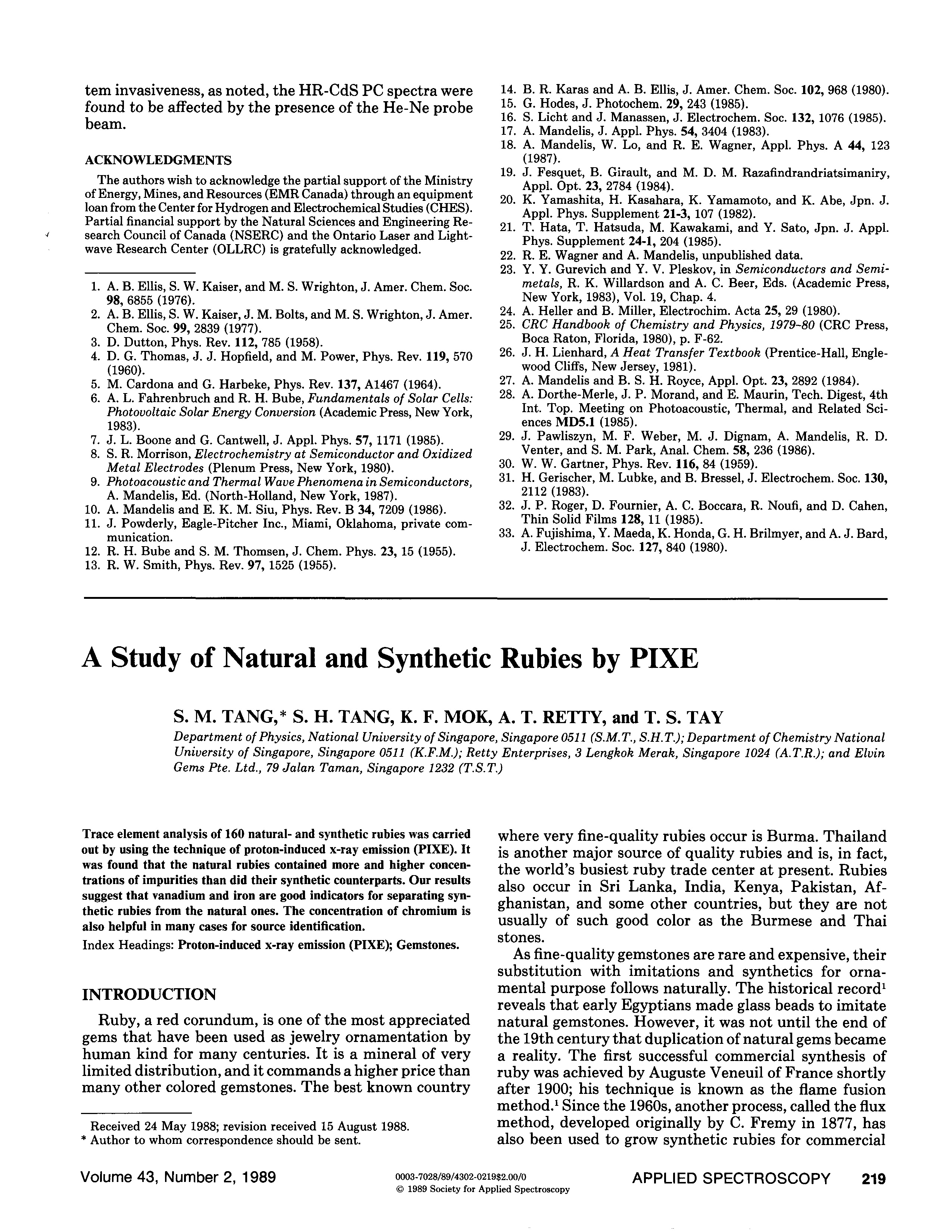
Tang, S.M., Tang, S.H., Mok, K.F., Retty, A.T. and Tay, T.S. (1989) A study of natural and synthetic rubies by PIXE. Applied Spectroscopy, Vol. 43, No. 2, pp. 219–223.
Record No. 5005  |  Sinkankas/Born No.: NIS/NIB  |  RWHL
Proton-induced X-ray emission (PIXE) is evaluated as a means of distinguishing natural from synthetic ruby and obtaining information about geographical or manufacturing source. Trace-element analyses were performed on 160 specimens comprising 133 natural rubies from Afghanistan, Burma, two Indian localities, Kenya, Sri Lanka, and Thailand, and 27 synthetics representing Chatham, Inamori, Kashan, Knischka, Ramaura, Seiko, and an unidentified flame-fusion source. Some natural origins were supported by characteristic inclusions, whereas others depended on supplier information because the specimens were insufficiently transparent for inclusion-based verification. Measurements used 2 MeV protons from a Van de Graaff accelerator, a silicon–lithium detector, and counting periods of 150 seconds per specimen. X-ray peak intensities were quantified by iterative least-squares fitting and calibration against standards. Natural rubies generally contained a greater variety and higher concentrations of impurities than synthetic specimens. Vanadium was undetectable in every synthetic analysed but was present in most natural stones, with exceptions among the Thai and Indian samples; the detection limit was approximately 20 ppm in the absence of titanium. Burmese rubies had the highest mean vanadium concentration, with one specimen containing 1800 ppm, while Thai rubies had the lowest mean. The authors therefore regard detectable vanadium as strong evidence of natural origin within the studied material, while stressing that its absence does not establish synthesis. Iron was generally low in synthetics, apart from the single Knischka specimen, and comparatively abundant in Thai rubies and some Sri Lankan stones. Kashan synthetics showed prominent titanium signals. Chromium concentrations varied substantially between source groups but were relatively restricted within several groups, suggesting supplementary value for source discrimination. Combined elemental profiles were more informative than any single concentration: iron, vanadium, and chromium together could, for example, help distinguish Sri Lankan from Thai material. The study concludes that PIXE offers rapid and useful trace-element characterisation, with vanadium and iron especially helpful for separating the examined natural and synthetic rubies. A larger reference database of specimens with established origins is recommended, although the authors caution that manufacturers could alter synthetic compositions and thereby weaken future chemical distinctions.

Fluid and mineral inclusions in corundum from gem gravels in Sri Lanka
Journal Article
Maesschalck, A.A., De and Oen, I.S. (1989) Fluid and mineral inclusions in corundum from gem gravels in Sri Lanka. Mineralogical Magazine, Vol. 53, December, pp. 539–545.
Record No. 4905  |  Sinkankas/Born No.: NIS/NIB  |  RWHL
Mineral and fluid inclusions were studied in seven gem corundums from gravels of three areas in Sri Lanka. All fluid inclusions are pure CO2. Microthermometry results on primary fluid inclusions suggest formation of corundum under granulite facies metamorphism (>630 ~ 5.5 kbar). Secondary fluid inclusions indicate different retrograde events of post-metamorphic cooling and uplift for different source areas.

Orangefarbene korunde aus Malawi

Orange corundum from Malawi
Journal Article
Henn, U., Bank, H. and Bank, F.H. (1989) Orangefarbene korunde aus Malawi. [Orange corundum from Malawi] Zeitschrift der Deutschen Gemmologischen Gesellschaft, Vol. 38, No. 4, pp. 164–166.
Record No. 4804  |  Sinkankas/Born No.: NIS/NIB  |  Language: In German  |  RWHL
Describes orange gem-quality corundum from the Chimwadzulu Hills of southern Malawi, near the Mozambique border. The material has refractive indices of nε = 1.763–1.765 and nω = 1.771–1.773, birefringence of 0.008, and specific gravity of 3.97–3.98. Microscopic features include twin lamellae, healed fractures and rounded doubly refractive mineral inclusions. Spectroscopic examination indicates that the orange coloration results from the superposition of absorptions due to Fe³⁺ and Cr³⁺. The authors contrast this mechanism with orange Sri Lankan corundum whose coloration may be produced by color centers.

Australian colour-changing sapphire

Journal Article
Brown, G. and Kelly, S.M.B. (1989) Australian colour-changing sapphire. Australian Gemmologist, Vol. 17, No. 2, pp. 47–48.
Record No. 4722  |  Sinkankas/Born No.: NIS/NIB  |  RWHL
G. Brown and S.M.B. Kelly describe an unusual apparent colour change in a natural sapphire from the Central Queensland sapphire fields, attributing the effect to the interaction of illumination with strongly zoned colour rather than to a true alexandrite effect. The specimen is a 2.03 ct oval brilliant that appears bluish in daylight and colour-corrected fluorescent light but purplish under incandescent illumination. Routine testing gives a hardness of 9, specific gravity of 3.98, refractive indices of 1.762 and 1.770, and birefringence of 0.008. Pleochroic colours are blue and reddish blue, the Chelsea-filter response is blackish, and no visible absorption features are reported. Examination also reveals a conspicuous orange patch within the otherwise predominantly blue sapphire. Microscopy establishes that this patch is discrete and closely associated with angular colour banding, readily seen when the stone is viewed obliquely through its crown. Rounded inclusions tentatively identified as zircon are scattered throughout the specimen and are surrounded by disc-like stress halos. Dust-like silk is prominent around both the angular zoning and the orange region. A particularly bright, irregularly outlined disc proves to be reflected light from a stress halo surrounding one of the presumed zircon crystals, rather than a separate reflective inclusion. Experimental illumination shows that the apparent colour depends on both the direction of incident light and its spectral composition. In blue-green-rich daylight, or when light passes through the major blue component of the stone, the sapphire appears blue. Under red-rich incandescent light, or when illumination is deliberately directed through the orange patch, it becomes reddish blue or dark purplish blue. Illumination from two separate directions can also produce a visibly bicoloured appearance. The authors locate the orange zone deep within the pavilion and regard it as the apparent control on the observed change. They therefore distinguish the specimen’s initially alexandrite-like appearance from a true alexandrite effect, explaining the observation in terms of differently coloured regions contributing unequally under changing lighting conditions. Photographs document its contrasting appearances, colour zoning, inclusions, and directional response. The report presents the stone as an exceptional example of colour-changing behaviour in Central Queensland sapphire while emphasizing the need to examine internal zoning and illumination geometry when interpreting such effects.

Star sapphire from Kenya

Journal Article
Barot, N.R., Flamini, A., Graziani, G. and Gübelin, E.J. (1989) Star sapphire from Kenya. Journal of Gemmology, Vol. 21, No. 8, October, pp. 467–473.
Record No. 4705  |  ISBN/ISSN: 221252  |  Sinkankas/Born No.: NIS/NIB  |  RWHL
Investigates the gemmological properties, inclusions, and causes of asterism in ten sapphire cabochons from northwestern Kenya, where commercially available rough began appearing on the local market in mid-1987. The material was reportedly collected from Quaternary surface sediments west of Lake Turkana, but its primary geological source remained unidentified. The samples ranged from blue through greenish-blue and greenish-yellow to dark brownish or nearly black, with transparency varying from moderately transparent to opaque. Eight displayed asterism, including six- and twelve-rayed effects, while two showed only concentrated silky patches. Four stones exhibited particularly sharp six-rayed stars with equally bright, narrow rays extending across the cabochons. Standard gemmological testing gave an approximate spot refractive index of 1.765, moderate pleochroism, and no reaction under long- or short-wave ultraviolet radiation or through the Chelsea filter. Most specimens showed iron-related absorption lines at 450, 460, and 470 nm; darker specimens instead showed a faint 470 nm line and a band between 450 and 460 nm. Microscopy, electron microprobe analysis, and X-ray diffraction distinguished cloudy areas, acicular features, silk, tubular cavities, transparent crystals, and feathers or healed fractures. The acicular systems responsible for asterism comprised microstriae chemically resembling the host corundum and needles attributed to rutile and hematite. Their crystallographic alignment and distribution governed the strength and completeness of the stars. Fine rutile needles contributed to silk, while hematite and other iron compounds were associated with darker colours. Additional inclusions comprised colourless tabular corundum crystals, brown prismatic crystals interpreted as rutile, and iron oxides or hydroxides filling channels and healed fractures. Angular blue colour zoning and inclusion arrangements were interpreted as records of successive growth episodes under changing physical and chemical conditions. Later fluid penetration introduced or redistributed iron compounds along fractures and cavities. Haloes around healed areas were tentatively linked to dehydration of iron hydroxides and formation of oxides during increasing temperature. The authors inferred high-temperature, high-pressure growth conditions and proposed that maximum crystallisation temperatures exceeded 500 °C. Although the finest stars closely resembled those of Linde synthetic star sapphires, straight angular growth zoning and characteristic iron absorption provided distinguishing evidence of natural origin. The study emphasises that an exceptionally regular star does not alone establish synthetic origin and that inclusion examination and spectroscopy are needed to characterise these Kenyan sapphires.

Notes from the laboratory [spectrophotometers]
spectrophotometers
Journal Article
Scarratt, K. (1989) Notes from the laboratory [spectrophotometers]. Journal of Gemmology, Vol. 21, No. 6, April, pp. 339–346.
Record No. 3252  |  ISBN/ISSN: 221252  |  Sinkankas/Born No.: NIS/NIB  |  RWHL
The expanding role of recording spectrophotometers in gem identification is illustrated through the development of the Gem Testing Laboratory of Great Britain’s equipment and several diagnostic case studies. Although the hand-held spectroscope remains valuable, its limitations become significant when precise wavelength measurements, absorption-band shapes, or information outside the visible region are needed to distinguish synthetic gems and treated natural material. Diamond absorption features provide an example: the H3 zero-phonon line at 503.2 nm and the H4 line at 496 nm require careful distinction because an H3 phonon sideband can be mistaken for the latter, potentially affecting interpretation of color origin. Cooling diamonds to approximately 120 K improves visual resolution, but recording instruments provide more complete spectral information. The laboratory initially acquired a Pye Unicam PU 8800 ultraviolet-visible spectrophotometer and subsequently added a Nicolet 510 Fourier-transform infrared spectrometer as infrared analysis became increasingly important. Guidance is given on interpreting absorbance and transmittance curves and relating recorded features to observations through a hand spectroscope. Examination of De Beers synthetic yellow to greenish yellow and brownish yellow diamonds combines ultraviolet luminescence, growth-sector zoning, metallic and dust-like inclusions, and infrared spectra. The examined synthetic stones show isolated-nitrogen absorption characteristic of type Ib diamond without aggregated-nitrogen features, whereas a natural yellow type Ib comparison stone also contains an aggregated-nitrogen peak. Infrared spectroscopy also identifies amber in mounted earclips and beneath artificial coatings on beads when conventional tests are restricted by mounting or surface treatment. The earclips display colored outer layers surrounding colorless interiors, consistent with heat treatment; the beads have an additional orange artificial coating over a yellow surface layer. A further case concerns a 5.80 ct diamond whose appearance varies among green, gray, and brown under different lighting. Its visible spectrum does not fit a straightforward conventional category. Strong blue-green phosphorescence following short-wave ultraviolet exposure and electrical conductivity suggest type IIb diamond, and comparison of its infrared spectrum with recorded reference spectra confirms that classification despite its atypical color. These examples demonstrate how instrumental spectroscopy complements microscopy and routine gemological observations, providing decisive information in cases where appearance or simpler tests are insufficient. The article argues that increasing identification challenges justify advanced instrumentation and that clearly presented spectral curves remain accessible and useful to practicing gemologists.

Gem Trade Lab Notes: Damaged Burma" ruby"
Journal Article
Hargett, D. (1989) Gem Trade Lab Notes: Damaged Burma" ruby". Gems & Gemology, Vol. 25, No. 4, Winter, pp. 240—241.
Record No. 3011  |  ISBN/ISSN: 0016626X  |  Sinkankas/Born No.: NIS/NIB  |  RWHL

Gem Trade Lab Notes: Ruby, natural color

Journal Article
Kane, R.E. (1989) Gem Trade Lab Notes: Ruby, natural color. Gems & Gemology, Vol. 25, No. 2, Summer, pp. 105–106.
Record No. 2980  |  ISBN/ISSN: 0016626X  |  Sinkankas/Born No.: NIS/NIB  |  RWHL

Exploring the Literature of Gemstone Inclusions

The literature of gemstone inclusions covers identification, origin determination, treatments, synthesis, mineral inclusions, fluid inclusions and other internal features of natural and synthetic gems.

For a curated selection of important sources, read Inclusions in Gemstones — Hyperion Literature Sources.

About Four Treasures

Four Treasures is Lotus Gemology's freely accessible gemological reference database. Use the full database to search by author, title or keyword and explore literature on gemology, mineralogy, jewelry and related fields. Where available, hyperlinks on titles may lead directly to the full publication.

Search the Complete Four Treasures Database