Gemstone Inclusions Bibliography

Gemstone Inclusions Bibliography

Explore books, articles and historical sources on inclusions in gemstones in Four Treasures, the Lotus Gemology reference database. This bibliography brings together matching records from the collection, with publication details, abstracts and cover images where available.

Gemstone Inclusion References

The list below matches the terms inclusion or inclusions in the database's searchable fields. Some references discuss inclusions as part of a broader subject.
841–860 of 1,377 matching references

Alexandrite, emerald, ruby, sapphire and topaz in a biotite-phlogopite fels from Poona, Cue District, Western Australia
Journal Article
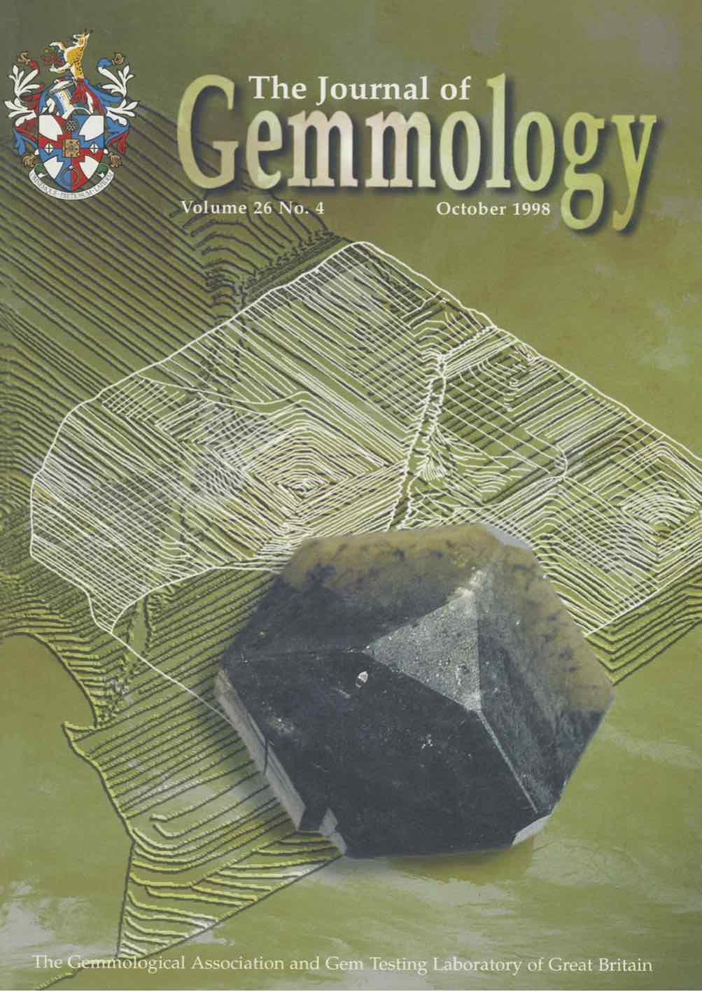
Grundmann, G. and Morteani, G. (1998) Alexandrite, emerald, ruby, sapphire and topaz in a biotite-phlogopite fels from Poona, Cue District, Western Australia. Australian Gemmologist, Vol. 20, No. 4, pp. 159–167.
Record No. 8040  |  Sinkankas/Born No.: NIS/NIB  |  RWHL
An unusual association of alexandrite, emerald, ruby, sapphire and topaz is described from Poona, near Cue in Western Australia, following field investigations in 1994. The discovery established the first documented occurrence of alexandrite-bearing bedrock in Australia. The minerals occur as preserved or fragmented porphyroblasts within biotite–phlogopite rocks belonging to a metasomatic blackwall sequence. Three types of emerald mineralization are distinguished: emerald associated with margarite and topaz; emerald accompanied by ruby, sapphire, topaz and alexandrite; and emerald associated with quartz and feldspar, with or without garnet and muscovite. The second assemblage, found in specimens from the dumps of the “Ruby shaft” near the Aga Khan Deep Mine, contains millimetre- to centimetre-scale bands interpreted as metasomatically altered greisens. Pink to violet fluorite, white micas and quartz accompany the gem minerals. Corundum commonly forms tabular crystals and radial or subparallel aggregates, with blue or colourless sapphire locally preserved within younger pink ruby. Many crystals display a deep blue core, a relatively colourless intermediate zone and a red outer zone, suggesting increasing chromium availability during growth. Alexandrite occurs as small prismatic crystals and characteristic star-like twinned aggregates, generally green to blue-green in daylight and blue to violet-red under incandescent illumination. Ruby and alexandrite fluoresce red under ultraviolet radiation, more strongly under long-wave than short-wave excitation, whereas colourless and blue corundum show no fluorescence. Corresponding colour zoning in corundum, chrysoberyl and beryl supports a multistage growth history involving changes in the supply of colouring elements. The proposed genetic model begins with fluorine-, beryllium- and rare-metal-bearing fluids from granites entering Archaean ultramafic rocks and producing greisens and mineralized veins. Subsequent regional metamorphism at upper greenschist to lower amphibolite facies promoted metasomatic reactions between these veins and their host rocks, forming corundum, alexandrite and emerald. During retrogression, topaz, corundum and chrysoberyl were partly or completely replaced by margarite, muscovite and locally emerald. Although the assemblage offers exploration potential, most crystals examined were small, fractured, inclusion-rich or altered, and little material suitable for faceting had been recovered. The economic prospects for ruby and alexandrite therefore remained uncertain.

The chemical signature of emeralds from the Campos Verdes-Santa Terezinha Mining District, Goiás, Brazil
Journal Article
Pulz, G.M., Silva, L.J.H.d.e.-R., Neto, L.S.B., Brum, T.M.M., Juchem, P.L., Santos, C.A., Pereira, V.P. and Silva, J.J. (1998) The chemical signature of emeralds from the Campos Verdes-Santa Terezinha Mining District, Goiás, Brazil. Journal of Gemmology, Vol. 26, No. 4, October, pp. 252–261.
Record No. 6063  |  ISBN/ISSN: 221252  |  Sinkankas/Born No.: NIS/NIB  |  RWHL
Emerald crystals from the Campos Verdes mining district have been analysed by inductively coupled plasma mass spectrometry (ICP-MS), Fourier transform infrared spectroscopy and Mössbauer 2+ 3+ spectroscopy. These gems contain measurable amounts of Fe , Fe , Cr, V, Sc, Ni and Zn; large-ion lithophile element (LILE, Ba, Sr, Rb, Pb) and high field strength element (HFSE, Zr, Y, Ti) contents are near or below the limit of detection. This chemical signature reflects the metasomatic reactions between the mineralizing fluids and the ultrabasic host-rocks of this gem deposit.

Lab Notes: Jadeite with inclusions of zircon
Journal Article
Koivula, J.I. and Moses, T. (1998) Lab Notes: Jadeite with inclusions of zircon. Gems & Gemology, Vol. 34, No. 1, Spring, p. 45.
Record No. 5496  |  ISBN/ISSN: 0016626X  |  Sinkankas/Born No.: NIS/NIB  |  RWHL
As the practice of bleaching and impregnating jadeite has become more common over the last few years, the amount of jadeite we test in the laboratory has increased signifi-cantly. During the microscopic exam-ination of one variegated green jadeite cabochon for possible evidence of treatment, we noted that the stone contained a number of small crystal inclusions, some of which reached the surface (figure 4). These surface-reaching crystals appeared to be somewhat harder than the surround-ing jadeite, and in reflected light they had a higher surface luster. With the recent acquisition of GIA’s Renishaw Raman Imaging Microscope System, we were able to examine two of these crystals nondestructively. By focusing the argon laser through the 50 x objective of the Leica targeting microscope and run-ning several continuous scans, we obtained clear spectra of the two “inclusions.” A computer search of our data base revealed that these spec-tra matched the spectrum for zircon. Visible characteristics of the inclu-sions (e.g., crystal form, color, trans-parency, and relative hardness) were consistent with this identification. This is the first time that we have encountered zircon crystals as inclu-sions in jadeite. Coincidentally, about a month prior to our discovery, a zir-con inclusion in a jadeite cabochon was also identified by means of the same technique at the Center for Gemstone Testing in Bangkok (Ken-neth Scarratt, pers. comm.).

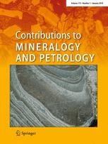
Models of corundum origin from alkali basaltic terrains: a reappraisal
Journal Article
Sutherland, L.F., Hoskin, P.W.O., Fanning, C.M. and Coenraads, R.R. (1998) Models of corundum origin from alkali basaltic terrains: a reappraisal. Contributions to Mineralogy and Petrology, Vol. 133, No. 4, pp. 356–372.
Record No. 5294  |  ISBN/ISSN: 107999  |  DOI: https://dx.doi.org/10.1007/s004100050458  |  Sinkankas/Born No.: NIS/NIB  |  RWHL
Corundums from basalt fields, particularly in Australia and Asia, include a dominant blue-green-yellow zoned magmatic suite (BGY suite) and subsidiary vari-coloured metamorphic suites. The BGY corundums have distinctive trace element signatures, with up to 0.04 wt% Ga₂O₃ and low Cr/Ga and Ti/Ga ratios (<1). Different melt origins for the BGY suite are examined based on inclusion and intergrowth mineralogy, petrologic associations, and tectonic setting. Over 100 analysed primary mineral inclusions include typical feldspars, zircon, Nb–Ta oxides, hercynite–magnetite, gahnospinel, rutile–ilmenite solid solution, calcic plagioclase, Ni-rich pyrrhotite, thorite, and low-Si, Fe-rich glassy inclusions. This expands upon prior surveys; New England, eastern Australia, in particular, shows the most diverse inclusion suite yet documented from basalt-hosted corundums (20 phases). Rare earth element patterns of zircon inclusions, intergrowths, and megacrysts are similar, with the exception of variable Eu depletion. Temperature estimates derived from magnetite exsolution textures, feldspar compositions, and fluid inclusion homogenization suggest corundum crystallization occurred at 685–900 °C. The overlap in Nb–Ta oxide compositions between corundum inclusions and those found in niobium–yttrium–fluorine (NYF) granitic pegmatites supports a silicate melt origin. The MELTS thermodynamic model was applied to amphibole–pyroxenite xenolith compositions from basalts to test the viability of corundum formation from mantle-derived melts. The calculations indicate corundum crystallization occurs at 720–880 °C and 0.7–1.1 GPa, producing residual feldspathic assemblages matching observed mineral inclusions and xenoliths. A model is thus proposed in which corundum in the BGY suite forms from low-volume silicate melts generated during partial melting of amphibole-bearing lithospheric mantle under the influence of magmatic plume activity.

An interdisciplinary approach to identifying solid inclusions in corundum: Thorite in a ‘Sri Lanka’ sapphire
Journal Article
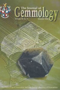
Carbonin, S., Sbrignadello, G. and Ajo, D. (1998) An interdisciplinary approach to identifying solid inclusions in corundum: Thorite in a ‘Sri Lanka’ sapphire. Journal of Gemmology, Vol. 26, No. 4, October, pp. 262–264.
Record No. 4732  |  ISBN/ISSN: 221252  |  Sinkankas/Born No.: NIS/NIB  |  RWHL
Carbonin, Sbrignadello, and Ajò demonstrate the value of combining independent analytical techniques to identify solid inclusions in corundum through a study of a commercially obtained blue sapphire containing thorite. The stone was provisionally assigned to a Sri Lanka type on the basis of an experienced supplier’s assessment, but reliable information establishing its geographical provenance was unavailable. Transmitted-light microscopy revealed at least two visually distinct inclusion types. A red, well-formed octahedral crystal associated with fingerprint inclusions was considered to be chromium-bearing spinel, a feature previously described in Sri Lankan sapphire. Photoluminescence measurements were undertaken with helium–neon laser excitation and subsequently with a more intense argon laser because the initial chromium-related emission from the host sapphire was weak and noisy. The principal identification problem concerned a second inclusion, approximately 45 µm long, whose habit, surrounding tension haloes, and fissures initially suggested metamict zircon. Because this grain lay close to the sapphire’s surface, the host was polished until the inclusion was exposed for electron microprobe examination. Energy-dispersive analysis unexpectedly detected no characteristic zirconium radiation. Instead, silicon and several diagnostic thorium emissions dominated the spectrum, supporting identification as thorite, ThSiO₄, whose crystal habit closely resembles that of zircon. The grain’s extremely small size prompted confirmation by an independent method. Low-background gamma spectrometry using an intrinsic germanium detector established the presence of natural thorium-232 through characteristic emissions from its radioactive daughter products, lead-212 and thallium-208, at 238.6 and 583.1 keV, respectively. The agreement between elemental microanalysis and nuclear spectrometry strengthened the thorite identification beyond what could be inferred from optical appearance alone. The authors emphasize that their complementary methods probe different physical processes, involving electronic and nuclear energy levels, and therefore provide independent support when a microscopic identification is uncertain. The study cautions against assigning zircon solely from crystal morphology and associated stress features, since another radioactive silicate can present a similar appearance. It also suggests that thorite should be considered among the inclusion species potentially relevant to sapphire provenance investigations, while the uncertain source of the specimen prevents its use as firm evidence of a Sri Lankan occurrence.

Spinel and garnet star networks: An interesting asterism in gems from Sri Lanka
Journal Article
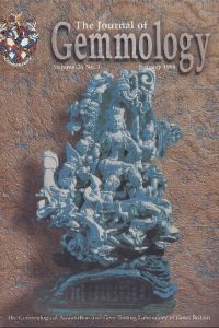
Kumaratilake, W.L.D.R.A. (1998) Spinel and garnet star networks: An interesting asterism in gems from Sri Lanka. Journal of Gemmology, Vol. 26, No. 1, January, pp. 24–28.
Record No. 4144  |  ISBN/ISSN: 221252  |  Sinkankas/Born No.: NIS/NIB  |  RWHL
Four-ray and six-ray star networks are reported in spinel and garnet from Sri Lanka. Details of their arrangement and orientations are discussed.

China company produces hydrothermal rubies
Journal Article
Anonymous (1998) China company produces hydrothermal rubies. Jewellery News Asia, No. 169, p. 50.
Record No. 2950  |  Sinkankas/Born No.: NIS/NIB  |  RWHL
Hydrothermal synthetic rubies marketed under the name “JL synthetic rubies” are available from Guangxi Jewelry Co. Ltd. in Guilin, China (Guangxi Province). The material ranges from red to orange-red in color, with some displaying purple hues. The synthetic rubies fluoresce strong red to long-wave ultraviolet radiation; the fluorescence is less intense to short-wave UV. Colorless “net-like” inclusions are reported. The synthetic rubies have R.I. values of 1.759–1.769, and an S.G. of 4.01. They are incorporated into gold rings, pendants, earrings, and bracelets. MM

Rubies and pink sapphires from the Pamir Range in Tajikistan, former USSR
Journal Article
Smith, C.P. (1998) Rubies and pink sapphires from the Pamir Range in Tajikistan, former USSR. Journal of Gemmology, Vol. 26, No. 2, April, pp. 103–109.
Record No. 2550  |  ISBN/ISSN: 221252  |  Sinkankas/Born No.: NIS/NIB  |  RWHL
Smith characterizes rubies and pink sapphires from marble-type deposits in the Turakuloma region of Tajikistan’s Pamir Mountains through examination of eight gemstones weighing 0.30–1.48 ct. Discovered in the early 1980s, these deposits had supplied only limited quantities to the international market. The best-known mine, Snejnaya, lies at approximately 3500 m, where severe weather restricts mining largely to June–August. The investigation combines standard gemological testing, immersion microscopy, identification of mineral inclusions by scanning electron microscopy with energy-dispersive analysis, optical and infrared spectroscopy, and semiquantitative X-ray fluorescence analysis. Samples are transparent, with saturated purplish-pink to purplish-red colours. Refractive indices of 1.761–1.762 and 1.770, birefringence of 0.008–0.009, specific gravity of 3.99–4.02 and uniaxial negative optic character are consistent with ruby and pink sapphire generally. Long-wave ultraviolet fluorescence is strong to very strong orange-red to red, while short-wave reactions are weaker. Internal growth structures indicate predominantly dipyramidal habits, with subordinate basal and rhombohedral faces; occasional colour zoning and rhombohedral twin lamellae are also recorded. Particularly useful microscopic features include columnar concentrations of short rutile needles and particles, highly resorbed calcite crystals arranged in roughly parallel rows, and small mineral or negative crystals accompanied by oriented equatorial thin films. Additional inclusions comprise titanite, zircon, rutile and plagioclase feldspar, together with healed fractures and negative crystals containing multiple fluid phases. Visible absorption is dominated by chromium, with broad bands near 405 and 550 nm and characteristic sharp lines. Infrared spectra show the expected corundum lattice absorptions but no detected mid-infrared features attributable to foreign minerals or structural hydroxyl groups. Analyses of five specimens identify chromium as the principal minor element responsible for colour, accompanied by titanium, vanadium, iron and gallium. Although the stones most closely resemble material from Quy Chau, Vietnam, their combined inclusion patterns appear distinctive and differ from features commonly encountered in several other ruby sources. Smith emphasizes the small sample population and the limited information available on production and reserves. At the time of writing, civil conflict, financial constraints and difficult access restricted exploitation, while fine faceted stones were generally 1.5 ct or smaller.

New Locality and a New Kind of Jadeite Jade from Guatemala: Rutile-Quartz-Jadeite
Conference Paper
Smith, D.C. and Gendron, F. (1997) New Locality and a New Kind of Jadeite Jade from Guatemala: Rutile-Quartz-Jadeite. Fifth International Eclogite Conference, Terra Nova 9, p. 35.
Record No. 28500  |  Sinkankas/Born No.: NIS/NIB  |  RWHL
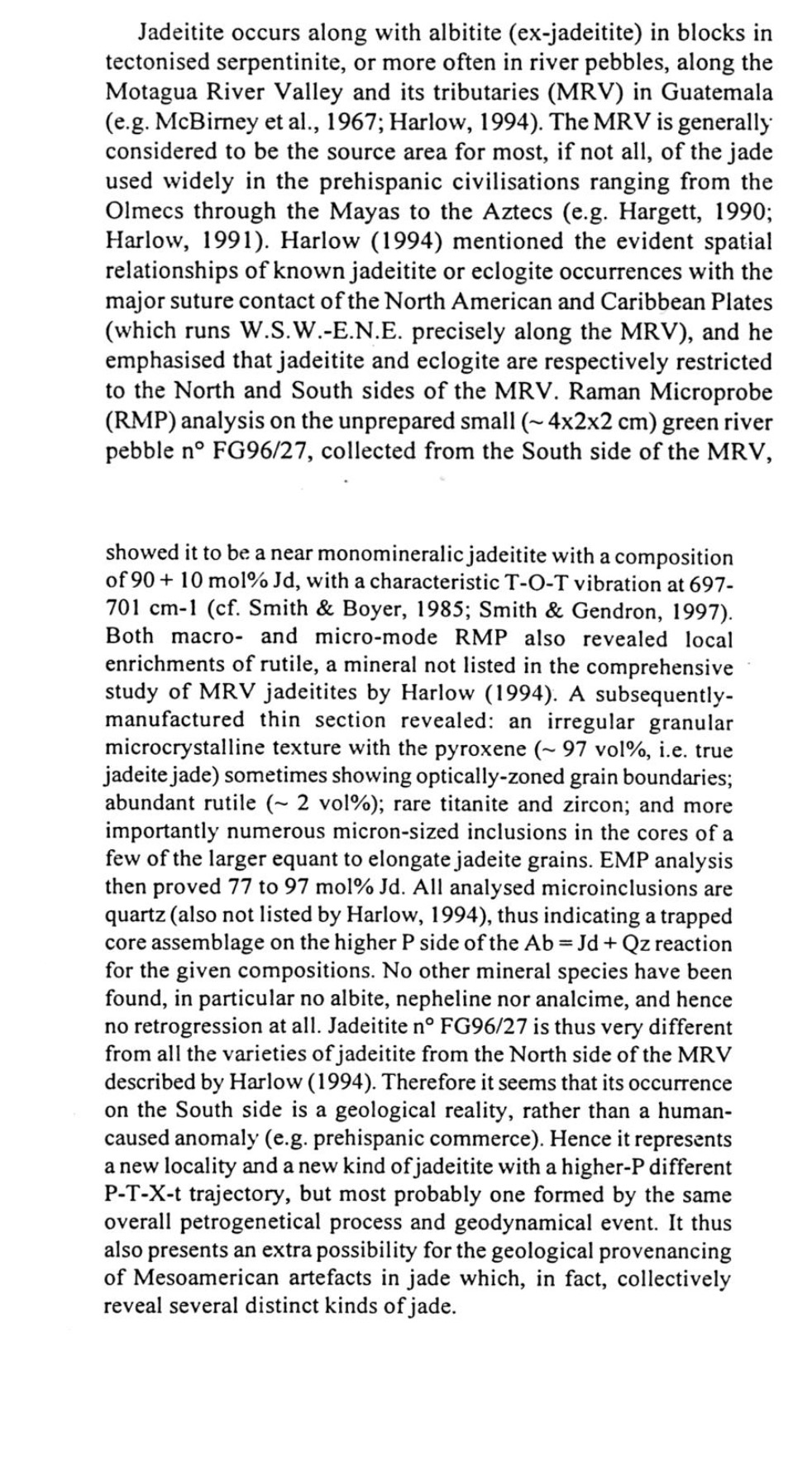
Jadeitite occurs along with albitite (ex-jadeitite) in blocks in tectonised serpentinite, or more often in river pebbles, along the Motagua River Valley and its tributaries (MRV) in Guatemala (e.g. McBirney et al., 1967; Harlow, 1994). The MRV is generally considered to be the source area for most, if not all, of the jade used widely in the prehispanic civilisations ranging from the Olmecs through the Mayas to the Aztecs (e.g. Hargett, 1990; Harlow, 1991). Harlow (1994) mentioned the evident spatial relationships of known jadeitite or eclogite occurrences with the major suture contact of the North American and Caribbean Plates (which runs W.S.W.-E.N.E. precisely along the MRV), and he emphasised that jadeitite and eclogite are respectively restricted to the North and South sides of the MRV. Raman Microprobe (RMP) analysis on the unprepared small (~4×2×2 cm) green river pebble n° FG96/27, collected from the South side of the MRV, showed it to be a near monomineralic jadeitite with a composition of 90 ± 10 mol% Jd, with a characteristic T-O-T vibration at 697–701 cm⁻¹ (cf. Smith & Boyer, 1985; Smith & Gendron, 1997). Both macro- and micro-mode RMP also revealed local enrichments of rutile, a mineral not listed in the comprehensive study of MRV jadeitites by Harlow (1994). A subsequently manufactured thin section revealed: an irregular granular microcrystalline texture with the pyroxene (~97 vol%, i.e. true jadeite jade) sometimes showing optically-zoned grain boundaries; abundant rutile (~2 vol%); rare titanite and zircon; and more importantly numerous micron-sized inclusions in the cores of a few of the larger equant to elongate jadeite grains. EMP analysis then proved 77 to 97 mol% Jd. All analysed microinclusions are quartz (also not listed by Harlow, 1994), thus indicating a trapped core assemblage on the higher P side of the Ab = Jd + Qz reaction for the given compositions. No other mineral species have been found, in particular no albite, nepheline nor analcime, and hence no retrogression at all. Jadeitite n° FG96/27 is thus very different from all the varieties of jadeitite from the North side of the MRV described by Harlow (1994). Therefore it seems that its occurrence on the South side is a geological reality, rather than a human-caused anomaly (e.g. prehispanic commerce). Hence it represents a new locality and a new kind of jadeitite with a higher-P different P-T-X-t trajectory, but most probably one formed by the same overall petrogenetical process and geodynamical event. It thus also presents an extra possibility for the geological provenancing of Mesoamerican artefacts in jade which, in fact, collectively reveal several distinct kinds of jade.

New emerald deposits from Southern India
Journal Article
Panjikar, J., Ramchandran, K.T. and Balu, K. (1997) New emerald deposits from Southern India. Australian Gemmologist, Vol. 19, No. 10, April–June, pp. 427–432.
Record No. 26923  |  Sinkankas/Born No.: NIS/NIB  |  RWHL
In the mid-1990s, emeralds were discovered embedded in the wall of a well at the village of Sankari Taluka, in southern India’s state of Tamil Nadu. Gemmological research into the chemistry, properties, and characteristic inclusions of these emeralds has revealed similarities between the properties of Sankari emerald and those of emerald from several previously described locations on the island of Madagascar. This finding suggests that Sankari emeralds may have been emplaced into the ancient supercontinent of Gondwanaland before India rifted from it some 200 million years ago.

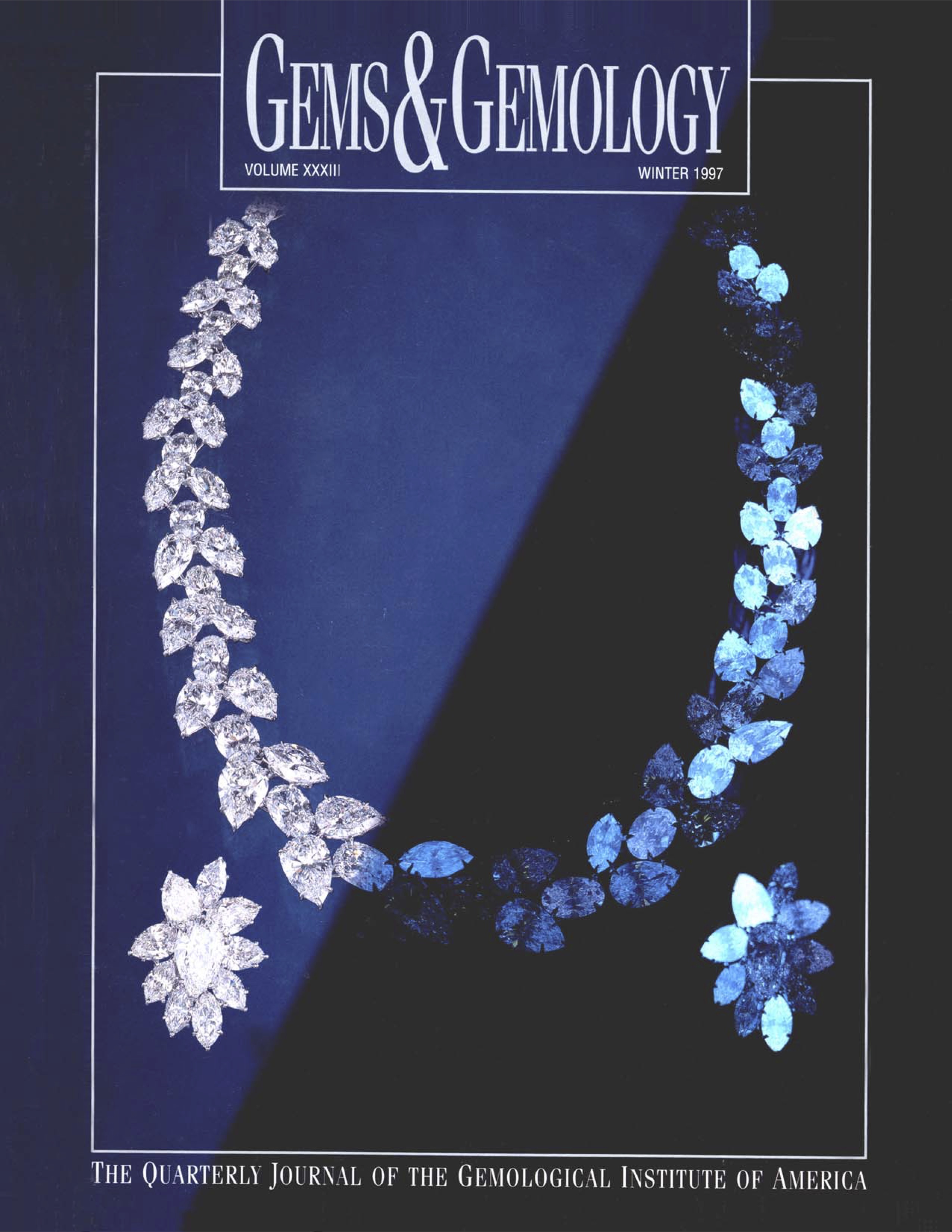
Characterization of alexandrite, emerald and phenakite from Franqueira (NW Spain)
Journal Article
Marcos-Pascual, C. and Moreiras, D.B. (1997) Characterization of alexandrite, emerald and phenakite from Franqueira (NW Spain). Journal of Gemmology, Vol. 25, No. 7, pp. 340–357.
Record No. 8041  |  ISBN/ISSN: 221252  |  Sinkankas/Born No.: NIS/NIB  |  RWHL
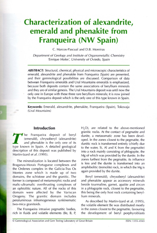
Marcos-Pascual and Moreiras examine the structural, chemical, physical and microscopic characteristics of emerald, alexandrite and phenakite from Franqueira in northwestern Spain, documenting an unusual beryllium-mineral association and assessing its gemological potential. Mineralization occurs in phlogopite-rich metasomatic rock developed where fluid-rich granitic pegmatites interacted with dunite. Beryllium supplied by the pegmatites combined with chromium derived from the ultramafic rock to produce emerald and alexandrite. The study employs single-crystal X-ray diffraction, electron microprobe analysis, refractometry, hydrostatic weighing, infrared spectroscopy and microscopy, supplemented by determinations of lithium, beryllium and water in emerald. Emerald is the most abundant beryllium mineral, forming light to medium green prismatic crystals up to 10 cm long. Most material is translucent and unevenly coloured, although distinct growth-related colour zoning was not observed. Chemical and structural data indicate predominantly octahedral substitution in the beryl structure, with channel alkalis contributing to charge compensation. Infrared spectra show that alkali-associated Type II water predominates over Type I water. Common inclusions comprise brown iron-rich phlogopite, negative crystals containing fluid phases, and healed fractures; less frequent mineral inclusions include quartz, pyrite, phenakite, alexandrite, ilmenite and anthophyllite. Previously reported fluid studies distinguish methane-bearing, carbon-dioxide-bearing and secondary saline aqueous inclusions. Alexandrite forms twinned crystals and skeletal inclusions in emerald, displaying a pronounced change from green or bluish-green in daylight to purplish-red under incandescent illumination, together with strong pleochroism. Abundant phlogopite inclusions and fractures limit the availability of good-quality cut stones. Phenakite is less abundant but can be transparent, colourless and suitable for faceting; its inclusions also include phlogopite and fluid-bearing healed fractures. Comparison with the genetically similar Tokovaja deposits in the Ural Mountains reveals broadly comparable alexandrite and phenakite properties, but differences in emerald colour, chemistry, infrared band intensities and inclusion morphology. Franqueira emeralds are generally paler because of lower chromium contents and lack several channel-like inclusion structures described in Uralian material. The authors relate these differences partly to the much smaller exposed extent and narrower geological diversity of Franqueira, while allowing that mineralization may continue underground. The study establishes a reference for identifying and understanding material from a locality vulnerable to depletion by collecting.

Gem News: Sapphires from northern Madagascar
Journal Article
Johnson, M.L. and Koivula, J.I. (1997) Gem News: Sapphires from northern Madagascar. Gems & Gemology, Vol. 33, No. 4, Winter, p. 305.
Record No. 6005  |  ISBN/ISSN: 0016626X  |  Sinkankas/Born No.: NIS/NIB  |  RWHL
Sapphires from northern Madagascar. In the past few years, several new and important sapphire deposits have been discovered in Madagascar. Dr. Margherita Superchi, of CISGEM, Milan, Italy, described a new occurrence at Ambondromifehy, in Antsiranana Province, in the northern part of the island. The sapphires found to date were alluvial, having been weathered from basaltic rocks that were emplaced intermittently during the period from the Upper Oligocene to the Pleistocene. The sapphires are thought to be xenocryts; that is, they did not crystallize from the basaltic magma, but simply were transported by it. They are associated with zircon, spinel, pyroxene, and olivine. Dr. Superchi and her colleagues (A. Donini, D. Muzzioli, and E. Roman) obtained detailed gemological data, chemical compositions, and optical and Raman spectra for 74 crystals (the largest being 182.59 ct) and 13 cut stones. Among the more interesting discoveries were inclusions of pyrochlore, zircon, and sodic plagioclase, which, with the chemical data (e.g., high gallium contents) confirm the “basaltic” association of these sapphires (see the earlier abstract on gem corundum).

Phase transformation of TiO₂ precipitates in sapphire (α-Al₂O₃) induced by the loss of coherency
Journal Article
Xiao, S.Q., Dahmen, U. and Heuer, A.H. (1997) Phase transformation of TiO₂ precipitates in sapphire (α-Al₂O₃) induced by the loss of coherency. Philosophical Magazine A, Vol. 75, No. 1, pp. 221–238.
Record No. 5529  |  Sinkankas/Born No.: NIS/NIB  |  RWHL
The first phase to precipitate in Ti-doped sapphire (Ti:α-Al₂O₃) single crystals annealed at 1300 °C in air is a high-pressure form of TiO₂ with the α-PbO₂ structure (α-TiO₂). The coherent precipitates form as oblate spheroidal plates with habit planes parallel to (0001). Because of a 5% lattice mismatch with sapphire, the small coherent α-TiO₂ precipitates are under an effective compressive stress, which stabilizes the high-pressure polymorph. During coarsening, the particles lose coherency by emitting a dislocation loop with a f(1̄101) Burgers vector, which reduces the stress on the particles and allows them to transform to the rutile structure. The orientation relationship between the sapphire matrix, α-TiO₂ and rutile has been determined and a lattice correspondence for the phase transformations established. Although the transformation from α-TiO₂ to rutile is purely structural, its rate is controlled by atomic diffusion; the increased atomic volume of rutile compared with α-TiO₂ is accommodated by expelling extra mass of Al₂O₃ into nearby interstitial dislocation loops and is associated with a change in the precipitate morphology.

A Raman microscope in the gemmological laboratory: First experiences of application
Journal Article
Hänni, H.A., Kiefert, L. and Chalain, J.P. (1997) A Raman microscope in the gemmological laboratory: First experiences of application. Journal of Gemmology, Vol. 25, No. 6, April, pp. 394–406.
Record No. 5371  |  ISBN/ISSN: 221252  |  Sinkankas/Born No.: NIS/NIB  |  RWHL
CMS: The Swiss Gemmological Institute (SSEF) in Basel has found its recent acquisition of a Raman microscope well worth the investment, to judge by this excellent account of its initial applications at this laboratory. Much more accurate, easier to use, and considerably less expensive than its predecessors, the Renishaw Raman microscope at SSEF easily fits on a desktop. It has enabled SSEF staff members to identify CO₂ inclusions in sapphire that are diagnostic of heat treatment, detect and identify fracture fillings in emerald, easily identify mounted gemstones, and characterize specimens with irregular surfaces. It can also distinguish component minerals in many aggregates. The method is nondestructive in all but a few well-documented conditions and works on all types of materials, except those having simple compositions and high symmetry, such as metals and alloys. Raman spectroscopy does require that the user be aware of its limitations and parameters; also, the lack of a large, published database means that each laboratory must obtain many of its own reference spectra from known samples. However, Raman spectroscopy clearly has earned a place of respect among the few high-tech laboratory techniques that have proved indispensable to gemological testing today.

Micro-PIXE analysis of trace element concentrations of natural rubies from different locations in Myanmar
Journal Article
Sanchez, J.L., Osipowicz, T., Tang, S.M., Tay, T.S. and Win, T.T. (1997) Micro-PIXE analysis of trace element concentrations of natural rubies from different locations in Myanmar. Nuclear Instruments and Methods in Physics Research B, Vol. 130, pp. 682–686.
Record No. 5368  |  Sinkankas/Born No.: NIS/NIB  |  RWHL
The authors analyzed 130 untreated rough rubies from nine metamorphic localities in Myanmar, including Mogok and Mong Hsu, using micro proton-induced X-ray emission (PIXE). The concentrations of chromium, titanium, vanadium, iron, copper, and gallium—the most abundant trace elements—were determined for a 200 × 200 μm area on each stone. In a unique approach, the authors used the trace-element mapping capability of the instrument to eliminate inhomogeneities, such as inclusions, from the analyses for the areas examined. The measurements revealed that, with the exception of gallium, there were large variations in the trace-element chemistry of rubies from the different localities, as well as for rubies from the same locality. Nevertheless, some consistent differences were also found. Using statistical analysis, including tree clustering and factor analysis, the authors determined that the origin of rubies from six of the nine localities could, indeed, be determined using this technique.

Inclusions in synthetic rubies and synthetic sapphires produced by hydrothermal methods (TAIRUS, Novosibirsk, Russia)
Journal Article
Peretti, A., Mullis, J., Mouawad, F. and Guggenheim, R. (1997) Inclusions in synthetic rubies and synthetic sapphires produced by hydrothermal methods (TAIRUS, Novosibirsk, Russia). Journal of Gemmology, Vol. 25, No. 8, October, pp. 540–561.
Record No. 5367  |  ISBN/ISSN: 221252  |  Sinkankas/Born No.: NIS/NIB  |  RWHL
This well-illustrated article examines (by SEM-EDX analysis) the inclusions in Russian hydrothermal ruby and sapphire manufactured by Tairus. Coatings on rough samples were found to be either boehmite or an unknown noncrystalline gel-like material. The following inclusions were observed in both rough and cut samples: solid inclusions of corroded copper (probably remnants of wire), isometric crystals and hexagonal platelets of copper, clouds and linear series of pinpoints of whitish reflective particles, tiny needles, and whitish particles (possibly alkali or calcium carbonates from the mineralizers). In pinkish red to red samples, the authors found fluid inclusion “fingerprints” or feathers, among which occur large three-phase (liquid-gas-mineral) inclusions associated with large tube-like negative crystals. The daughter minerals in the inclusions were identified (by crossed polarizers and heating/freezing [H/F] experiments) as carbonates. H/F experiments also revealed that the liquid phase is predominantly H₂O. By contrast, three-phase inclusions in natural rubies are often CO₂-rich with daughter minerals of diaspore, graphite, or mica. Of perhaps greatest interest to the practicing gemologist, the authors describe a simple gemological H/F test to distinguish between natural and synthetic hydrothermal rubies on the basis of three-phase inclusions. The copper inclusions also are indicative of synthetic origin.

Gemmological features of rubies and sapphires from the Barrington volcano, Eastern Australia
Journal Article
Webb, G. (1997) Gemmological features of rubies and sapphires from the Barrington volcano, Eastern Australia. Australian Gemmologist, Vol. 19, No. 11, pp. 471–475.
Record No. 2955  |  Sinkankas/Born No.: NIS/NIB  |  RWHL
Ruby is rare in Australia, but in the Barrington area of New South Wales, ruby and pink sapphire form up to 50% by weight of the gem corundum concentrate. These stones have Cr₂O₃ contents of 0.60–1.42 wt.% and FeO contents of 0.44–0.75 wt.%. They typically range from 2 to 6 mm in size and contain inclusions of spinel and liquid-filled fractures. Polysynthetic twinning is common and is often associated with inclusions of boehmite needles. On the basis of trace-element content, the Barrington corundum is divided into two distinct suites: one related to ruby and the other typical of eastern Australian sapphires. The ruby-containing corundum suite originated in a metamorphic environment, whereas the non-ruby suite has a magmatic origin. Both suites were brought to the surface by basaltic volcanism.

Le saphir padparadscha

The padparadscha sapphire
Journal Article
Notari, F. (1997) Le saphir padparadscha. [The padparadscha sapphire] Revue de Gemmologie A.F.G., No. 132, pp. 24–27.
Record No. 2538  |  Sinkankas/Born No.: NIS/NIB  |  Language: in French  |  RWHL
Notari investigates the physical and chemical characteristics of padparadscha sapphire to clarify inconsistent definitions of its colour and identity. Drawing on historical research, observations in Sri Lanka, professional opinions, and laboratory analyses, he defines its characteristic colour as orangey pink of pastel to medium saturation. Four appearances are distinguished: “original,” “antique,” “induced,” and “deceptive,” with only the first two accepted as genuine padparadscha. The others owe their appearance to treatment or yellowish iron oxides in fractures. Chemical and spectroscopic investigations examine the contributions of chromium, iron, and incompletely understood colour centres, together with blue growth zoning and sunlight-induced colour changes. Raman analysis identifies native sulphur within a multiphase inclusion containing diaspore, graphite, rutile, and carbon dioxide. The article discusses heating and irradiation treatments, their detection, and diagnostic features of melt-grown, flux-grown, and hydrothermal synthetics. Notari concludes that padparadscha is a colour variety rather than a geographical designation, while noting that only Sri Lankan production then regularly supplied stones matching the complete suite of characteristics described.

Charakterisierung von Saphiren mit Hilfe von Flüssigkeitseinschlüssen
Characterization of sapphires using fluid inclusions
Journal Article
Bruder, B. (1996) Charakterisierung von Saphiren mit Hilfe von Flüssigkeitseinschlüssen. [Characterization of sapphires using fluid inclusions] Zeitschrift der Deutschen Gemmologischen Gesellschaft, Vol. 45, No. 2, pp. 47–54.
Record No. 29619  |  Sinkankas/Born No.: NIS/NIB  |  Language: in German  |  RWHL
Examines fluid inclusions in sapphires from Myanmar, Kashmir, Sri Lanka, Madagascar, and Rwanda using microthermometry and Raman spectroscopy. The fluids consist of carbon dioxide, while diaspore daughter crystals indicate that water was formerly present. Four inclusion types are distinguished by their morphology, occurrence, and fluid density. Primary inclusions form well-developed, approximately equidimensional negative crystals containing high-density fluid. Secondary inclusions include short prismatic cavities within well-preserved healed fractures, together with two further types associated with incompletely healed fractures. Differences in the densities of primary fluid inclusions allow sapphires from Sri Lanka and Kashmir to be distinguished from those of the other localities examined, demonstrating the potential of fluid-inclusion analysis as an aid to sapphire characterization.

Gem News: “Golden” tourmaline from Kenya

Journal Article
Johnson, M.L. and Koivula, J.I. (1996) Gem News: “Golden” tourmaline from Kenya. Gems & Gemology, Vol. 32, No. 2, Summer, pp. 135–136.
Record No. 26763  |  Sinkankas/Born No.: NIS/NIB  |  RWHL
Tourmaline from a recent find in Kenya, referred to as “savannah” or “golden” tourmaline, has attracted attention for its distinctive coloration. Gemologist Yianni Melas submitted two rough samples for examination: a 4.50 ct transparent brownish orangy yellow piece with green areas, and a 3.06 ct crystal section showing a brownish orangy yellow core with a green rim. The broken crystal section displayed only prism faces, lacking striations parallel to the c-axis. Gemological properties recorded include pleochroism from brownish orange to yellow in the yellow regions and dark green to light yellowish green in the green areas, uniaxial optic character, refractive index of 1.630–1.650 with a birefringence of 0.020 (on the green rim), and specific gravity of 3.05. Under long-wave UV, the stones were inert, but showed moderately chalky yellow fluorescence under short-wave UV, stronger in the green rim. Spectroscopic analysis showed general absorption to ~480 nm, with additional bands between 590–630 nm and lines at 650 and 680 nm in the green rim. Inclusions observed included anhedral crystals, needles or tubes, two-phase inclusions, and hollow tubes. EDXRF spectroscopy revealed Mg, Al, Si, K, Ca, Ti, Fe, Zn, Ga, and Sr in both samples, with one showing a small Mn peak. Chromium was detected in the green rim, consistent with the observed color-filter and absorption data, indicating Cr as the chromophore for the green. Reported sources for this material include the Kasigau Mine in Tsavo West National Park, the Mangari area in the same park, and M’gama Ridge south of the Taita Hills in southeastern Kenya.

Exploring the Literature of Gemstone Inclusions

The literature of gemstone inclusions covers identification, origin determination, treatments, synthesis, mineral inclusions, fluid inclusions and other internal features of natural and synthetic gems.

For a curated selection of important sources, read Inclusions in Gemstones — Hyperion Literature Sources.

About Four Treasures

Four Treasures is Lotus Gemology's freely accessible gemological reference database. Use the full database to search by author, title or keyword and explore literature on gemology, mineralogy, jewelry and related fields. Where available, hyperlinks on titles may lead directly to the full publication.

Search the Complete Four Treasures Database